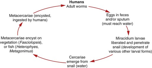
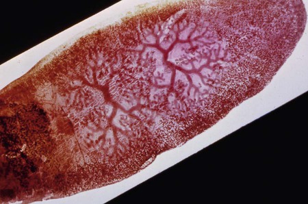
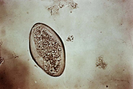
1. List the clinically significant intestinal trematodes. 2. Describe the general life cycle of the intestinal trematodes and identify the life cycle stage infective for humans. 3. Describe the diagnostic methods used to identify intestinal trematodes. 4. Explain the pathogenesis of intestinal trematode infections. 5. List the drugs of choice for intestinal trematode infections. 6. Describe the environment where intestinal trematodes are found, the route of transmission, and preventive measures. The adult worms are located in the small intestine, where they lay eggs that may be embryonated or remain unembryonated until they are shed from the body via feces. The egg continues developing after reaching the water, and a ciliated, free-swimming miracidium larva is released. The miracidium enters a snail host and develops into a redia (cylindrical larvae), followed by development into tailed cercariae. The cercariae emerge from the snail and encyst as a metacercariae (encrusted larvae) on water plants or fish. A human host ingests raw or undercooked plants (Fasciolopsis buski) or fish (Heterophyes heterophyes, Metagonimus yokogawai) containing the metacercariae, which exycyst in the intestinal tract, attach, and mature into adults (Figure 56-1). The adults of F. buski have an elongated shape and range from 20 to 75 mm long to approximately 8 to 20 mm wide (Figure 56-2). They have an oral sucker at the anterior end and a ventral sucker located about midway to the posterior end. The eggs, which are indistinguishable from those of Fasciola hepatica (Figure 56-3), are oval and elongated, transparent, and yellow-brown with an operculum (lid) at one end, and they range in size from 130 to 140 µm long to 80 to 85 µm wide and may be unembryonated.
Intestinal Trematodes
Fasciolopsis Buski
General Characteristics
![]()
Stay updated, free articles. Join our Telegram channel

Full access? Get Clinical Tree


Basicmedical Key
Fastest Basicmedical Insight Engine