Immunochemistry and Molecular Biology in Cytological Diagnosis


Immunochemistry and Molecular Biology in Cytological Diagnosis


Ronald A. DeLellis

Rana S. Hoda


As we develop methods for extrapolating the secrets previously locked within the individual cells, it becomes evident that the cells were talking all along; we just did not know how to listen.


The past several decades have witnessed the development of a remarkable array of methodological advances in the biomedical sciences. One of the most successful of these approaches has been immunocytochemistry. Methodologies employing antibodies as specific probes for the visualization of cell and tissue bound antigens have literally revolutionized the practice of pathology (Taylor, 1978; DeLellis et al, 1979; Taylor, 1994; Wick et al, 2001). The application of these methods has permitted the extension and expansion of morphology by means of increasingly more sensitive and specific markers that can be visualized in single cells and in tissue sections. Experience gathered over the past three
decades has indicated that the success of any immunohistochemical procedure depends on multiple factors, such as the antigen preservation, the specificity and avidity of the primary antibody and the sensitivity of the detection system. These factors are interrelated; reliable and reproducible results can be achieved only when each variable is optimized.

Immunochemistry on cytologic material has great potential utility as recognized by Nadji and others in the early to mid-1980s; however, problems with scanty cell samples, high levels of nonspecific background staining and falsepositive and false-negative reactions have limited the widespread application of this technique (Nadji, 1980; Chess and Hadju, 1986). Improved cytological collection methods, together with advances in immunocytochemical technology, including the development of new monoclonal antibodies and more sensitive detection methods, have led to greater degrees of sensitivity and specificity in cytological preparations (Ramaekers et al, 1988; Flens et al, 1990; Domagala and Osborn, 1992; Dabbs et al, 1995; Abati et al, 1998; Bedrossian, 1998; Dabbs and Wang, 1998; Fetsch and Abati, 1998). Shield et al (1996) analyzed the utility of immunocytochemical methods in cytology over a 20-month period and demonstrated that this approach was helpful in approximately 75% of cases. In body fluids, staining was performed most commonly for the distinction of mesothelial cells from metastatic malignancies. Immunocytochemistry was helpful in 82% of these cases with the preliminary diagnosis confirmed in 64%, refined in 8% and revised in 10%. In fine needle aspiration (FNA) samples, staining was helpful in 69% of cases, resulting in refinement of the diagnosis in 55% and confirming the preliminary diagnosis in 14%. In one case, these studies led to a revision of the original interpretation. In cytology, the practical utility of immunocytochemistry includes characterization of poorly differentiated neoplasms, differentiation of primary from metastatic tumors, determination of the sites of origin of metastatic lesions and prognostic assessments.

The purpose of this chapter is to provide an overview of immunocytochemical methods, their applications and limitations in different types of cytological preparations and their contributions to the understanding of neoplastic disorders. In addition, this chapter highlights molecular techniques that are of value in the analysis of cytological samples. For additional details including use of sophisticated techniques, such as linker and polymer-based staining, the reader is referred to the textbook by Dabbs (2002).*


PREPARATORY METHODS AND ANTIGEN PRESERVATION

Numerous preparatory techniques have been utilized for the immunochemical evaluation of cytological preparations (Table 45-1) (Sherman et al, 1994; Hunt et al, 1998; Mitteldorf et al, 1999). When sufficient material is available, formalin-fixed paraffin miniblocks and cell blocks offer a number of advantages since fixation and subsequent processing are essentially identical to those used for tissue sections (Domagala et al, 1990; Bellotti et al, 1997; Fowler et al, 1998). Moreover, antibody panels and multiple positive and negative controls can be applied to the same material. When the amount of cellular material is limited, other preparations including direct smears (unstained or previously stained), cytocentrifuge preparations, cells collected on filters or in liquid fixatives and cell transfers can be used (Table 45-1) (Sherman et al, 1994; Abendroth and Dabbs, 1995; Leung and Bedard, 1996; Gill, 1998).


Fixation

The choice of appropriate fixative plays a critical role for the optimal preservation of the antigen of interest and numerous standard and novel fixatives have been used for this purpose (Okuyama et al, 1996). Prompt fixation is necessary to minimize diffusion and extraction of soluble antigens and to preserve morphological integrity. Delays in fixation may lead to a loss or significant reduction of immunoreactivity due to diffusion of antigens from their intracellular sites and autolytic processes. Autolysis also results in nonspecific binding of antibodies to unrelated antigenic determinants. Despite this fact, a variety of antigens can be localized, even in necrotic samples of both histological and cytological preparations, provided that appropriate controls are used (Judkins et al, 1998; Marzec et al, 2001). Buffered neutral formalin (4% formaldehyde) is compatible with the localization of many antigens, both in histological and cytological samples. The popularity of this fixative is based on many factors, including its low cost, ease of preparation and its preservation of morphological detail (Werner et al, 2000). The basis of formalin fixation is the formation of hydroxyl-methylene type linkages between protein end groups which increase with the length of fixation time. An additional mechanism is the formation of coordinate bonds for calcium ions (Werner et al, 2000). Prolonged fixation in formalin, however, often leads to reduced immunoreactivity of many antigens, primarily because of the steric hindrance resulting from the formation of extensive crosslinkages. Crosslinks may be formed between two parts of the antigen resulting in masking of the epitope or between two or more different molecules. Such linkages may also directly affect the epitope itself (Werner et al, 2000). In the past, several approaches, including washing of sections with buffer or treatment with proteolytic enzymes, were used in restoring the reactivity of many epitopes. More recently, antigen retrieval methods based on the use of microwave heating of sections have permitted the localization of most antigens of interest to the pathologist as discussed in the next section.

In cell blocks prepared from previously alcohol-fixed spun sediment, fixation in formalin of less than 24 hours
may result in a mixture of formalin fixation and ethanol fixation, the latter resulting from tissue processing in graded ethanols. Brief formalin fixation will result in cross-link formation only at the periphery of the block while the center will be fixed in ethanol. As a result, sections prepared from such blocks may show variable staining that is more or less intense staining at the center or periphery, depending on the reactivity of the antibody and the retrieval procedure employed (Werner et al, 2000).


TABLE 45-1 IMMUNOCYTOCHEMISTRY USING DIFFERENT TYPES OF CYTOLOGICAL PREPARATIONS


Preparation


Fixative


Advantages


Disadvantages


Cell blocks (occasionally small tissue fragments retrieved from aspirated material)


Formalin fixation is usual; however, other fixatives may be used depending on nature of antigen


– Staining conditions virtually identical to standard histologic preparations
– Good cellular morphology and nuclear detail
– Serial sections may be prepared for use with antibody panels
– Standardization of retrieval methodology


– Requires relatively large amount of material
– Cannot be used with highly labile antigens
– Processing time is longer than with direct smears
– Effects of prolonged storage are unknown


Direct smears


Alcohol or formalin fixation


– May be the only material available for analysis
– Cellular areas can be subdivided or cells can be peeled off for multiple antibody tests
– Previously stained slides can be used with or without prior decolorization


– Limited material may compromise use of antibody panels and appropriate controls
– Cell loss may occur during staining
– Three dimensional cell groups may “trap” antibodies leading to nonspecific staining
– Mechanical smearing process may lead to disruption of cells and leakage of antigens
– Requires large quantity of antibodies and other reagents
– Potential for high background staining due to blood and necrotic material


Cytocentrifuge preparations


Alcohol or formalin fixation


– Lack of background staining
– Ease of interpretation since cells are concentrated in a small area
– Multiple slides can be prepared
– Relatively small amounts of antibodies or other reagents are used


– Significant amount of cellular material may be lost during cytocentrifugation process
– Lack of reproducibility if processing is not well controlled


Filters


Alcohol or formalin fixation


– Provides optimal recovery of cells


– High non-specific background staining


Thin Prep (Cytyc Corp., Marlborough, MA)
SurePath (TriPathology Imaging, Burlington, NC)


Proprietary fixatives


– High cell yield in initial preparations
– Clean background and absence of air-drying effect
– Multiple slides and cell blocks from residual material
– Relatively small amount of antibody required
– Single preparations can be subdivided


– Decreasing cell yield with sequential preparations
– High cost
– Certain markers (e.g., lymphoid markers) may be difficult to demonstrate


In addition to formalin, other fixatives have been employed for the demonstration of cell and tissue bound antigens, including the mercury containing fixatives such as Zenker’s and B5 (formol sublimate), picric acid containing fixatives (Bouin’s and Zamboni’s) and alcohol-based fixatives such as Carnoy’s fluid (alcohol, chloroform, acetic acid) (see Chap. 44). Additionally, ethanol, methanol and acetone have been used as primary fixatives for a variety of immunochemical applications. Alcohol and acetone are
precipitating or coagulating fixatives rather than cross-linking reagents; accordingly, retrieval procedures are not generally useful in restoring reactivity following fixation with these reagents. However, for some antigens a retrieval step may help in such preparations provided that the cells have been collected on adhesive slides.

It should be noted that fixatives can markedly alter the reactivity of antigens to antibodies since exposure patterns of epitopes can differ. As noted by Willingham et al (1999), most of the epitopes (the parts of antigens reacting with antibodies; for full explanation, see below) that are detectable by monoclonal antibodies are conditional in the sense that they are reactive only under certain conditions. For example, Josephsen et al (1999) have demonstrated that a monoclonal antibody directed against vimentin reacts with a novel epitope on an unrelated protein (amelogenin), but this phenomenon occurred only after fixation in 1% glutaraldehyde or 4% paraformaldehyde plus 0.1% glutaraldehyde but not with 2.5% glutaraldehyde. Blocking experiments demonstrated that the amelogenin antibody competed with the vimentin antibody.

The ideal fixative for immunocytochemistry of cytological preparations should be convenient to use and should provide both optimal antigen preservation and cellular morphology. Suthipintawong et al (1996, 1997) examined the effects of a large number of fixatives including acetone, acetone/methanol, acetone formalin, glutaraldehyde, ethanol, methanol and formol saline on antigen preservation. They concluded that fixation of air-dried smears in 0.1% formol saline (normal saline 1,000 ml and 40% formalin 2.5 ml) overnight at 27°C followed by 10 minutes fixation in 100% ethanol produced the most consistent results. Air drying is an important step since it minimizes the cell loss that inevitably occurs when fresh smears are immersed directly into fixative. As noted by Leong et al (1999), postfixation in ethanol is not essential for the preservation of immunoreactivity but improves the cytomorphological features. Formol saline produces complete hemolysis of background erythrocytes and removal of background proteinaceous fluid. Moreover, immunoreactivity can be further enhanced by microwave-induced epitope retrieval following formalin fixation. For air dried smears, adhesive-coated glass slides were not required. The smears could be kept at room temperature for at least 7 days and at − 70°C for 5 weeks without loss of immunoreactivity as air dried smears or after fixation in formal saline.

Abati et al (1998) have recommended short term storage of unfixed air dried cytological preparations at 4°C and long term storage at − 20°C in desiccant. Following removal of the slides from the refrigerator or freezer, it is important to allow the containers to equilibrate to room temperature prior to opening. They further recommend that the choice of fixative should be tailored to the particular antigen under study. For hematopoietic markers, they recommend 10 minutes fixation in acetone while a 1:1 mixture of methanol and absolute ethanol is used for nonhematopoietic markers. They recommend fixation in 3.7% buffered formalin for 15 minutes for those markers which have a nuclear localization (e.g., steroid receptors, p53). Freeze drying and freeze substitution methods have also been used for preservation of antigenicity in cytological materials (Takahashi et al, 1996).


Antigen Retrieval Methods

Suboptimal preservation of antigens is a major factor leading to lack of staining consistency in immunohistochemistry and, even more so, in immunocytochemistry. The effects of formalin fixation can be reversed in part by prolonged washing of cells and tissues in water or buffer. Proteolytic enzyme digestion of formalin fixed samples has proven to be an invaluable approach for the unmasking of some formalin sensitive epitopes. However, some antigens are highly susceptible to the effect of proteolysis and may be completely destroyed even after relatively brief periods of enzyme treatment. Proteolytic enzyme induced unmasking most likely results from breakage of formaldehyde induced cross links in the antigen itself, with exposure of cryptic epitopes or hydrolysis of adjacent macromolecular complexes that may have covered or masked the epitopes of interest. Optimization of the time of proteolysis appears to be more crucial to the success of this approach than the particular enzyme used. Generally, tissues fixed for prolonged time periods require the most extended periods of enzyme digestion.

A major advance in the retrieval of formalin sensitive epitopes involves the use of moist microwave heating of tissue samples. Pioneered by Shi et al (1991), antigen retrieval using microwave heating of sections immersed in 1% zinc sulfate or saturated lead thiocyanate resulted in significant increases in immunostaining with a high proportion of tested monoclonal antibodies. The precise mechanism by which microwave heating restores immunoreactivity, however, remains unknown. One possibility is that heating can lead to disruption of formalin induced bonds between proteins and calcium ions (Mogan et al, 1994). The most commonly used method utilizes microwave heating in 0.01 mol/L citrate buffer at pH 6.0. Other heating approaches include the use of pressure cookers and rice steamers. This method has also been applied successfully to cytological samples (Reynolds et al, 1994; Schmitt et al, 1995).

Since the types and lengths of fixation may differ widely a “test battery” approach has been proposed (Shi et al, 1996; Taylor and Shi, 2001). This approach is based on the heating conditions and the pH of the retrieval solution. According to Shi et al (1996), a maximal retrieval level that shows the strongest staining intensity can be established by using this test battery. The use of an optimal retrieval protocol permits comparable staining results for a variety of archival tissues that have been fixed in formalin for widely different time periods. The heat induced antigen retrieval methods also have potential pitfalls. Since most monoclonal antibodies react with epitopes containing three to eight amino acids, retrieval methods could potentially expose unwanted epitopes of identical sequence in other antigens. This could lead to unexpected cross reactions and the problem of false positive reactions as discussed previously. Heat induced methods have also been used for the retrieval
of DNA and RNA from archival samples. Heat induced antigen retrieval may also result in enhanced reactivity of endogenous biotin. This is particularly problematic in mitochondria-rich cells. When using an avidin biotin-based detection system, therefore, it is critical that endogenous biotin be blocked with avidin prior to staining (Rodriquez-Soto et al, 1997) or that a non-avidin—biotin system be used.


ANTIBODIES AND IMMUNOCYTOCHEMICAL METHODS

Antibodies are immunoglobulin molecules that are produced by B cells. They consist of a pair of light chains (kappa or lambda) and a pair of heavy chains (gamma, alpha, mu, delta, or epsilon) that are joined together by disulfide bonds (Listrom and Fenoglio-Preiser, 1992; Abbas et al, 2000). IgG has two antigen binding sites, each of which is able to bind with one molecule of the antigen. The antigen binding sites are located in the Fab region of the molecule and are formed by the N-terminal ends of the light and heavy chain pairs. Antigen binding occurs in that region of the Fab fragment that contains the variable domains of both the heavy VH and light VL chain gene sequences. Specificity is determined by the precise amino sequences of the variable domains. The Fc portion of the antibody molecule contains only heavy chains and accordingly does not bind to the antigen. However, the Fc portion of the molecule can react with receptors on the surfaces of many cells and can lead to non-specific binding of antibodies to cells. Complement binding sites are also present on the Fc portion of the immunoglobulin molecule and are another potential source of non-specific staining in immunohistochemical formats.

An antibody binds only to a specific portion of an antigen which is called a determinant or epitope. With phospholipids or complex carbohydrates, the antigenic determinants are a function of the covalent structure of the macromolecule. Noncovalent folding of protein macromolecules also contributes significantly to the formation of antigenic determinants. Epitopes are generally small and consist typically of three to eight amino acids. Those epitopes formed by adjacent amino acids in the molecule are termed linear or continuous determinants. If continuous epitopes are present on the surface of the native protein antigen, they will be able to react with the corresponding antibody. More often, continuous epitopes may be inaccessible to the antibody in their native conformation and will become reactive only when the antigen is denatured. Discontinuous epitopes are formed by amino acid sequences from separated portions of the molecule that are juxtaposed only in their native folded state. Such epitopes are typically lost upon denaturation of the molecule. This phenomenon explains, at least in part, discrepancies between the results of antibody assays using immunohisto- or cytochemical and Western analyses (Willingham, 1999).

Although the two binding sites of an immunoglobulin molecule could bind to two separate epitopes on the same or separate molecules of the antigen, probably only one site is bound to antigens in tissues. An antigen with several determinants could bind several molecules of antibody or could bind several antibodies of differing specificities directed against two or more different determinants present in the same antigen. Because of the relatively small number of amino acids in an epitopic sequence, cross reactivities of antibodies may occur in unrelated proteins and are a continual source of concern in immunochemical assays. A conformational change in an epitope, resulting from changes in pH, type and duration of fixation, temperatures of processing and exposure to solvents may, therefore, have a profound impact on the reactivity of the antibody with the epitope.

Although many early immunochemical studies were performed with polyclonal antisera, more recent studies have utilized monoclonal antibodies (Taylor and Cote, 1994). Polyclonal antisera are prepared by injecting the immunogen of interest into an animal with the subsequent stimulation of multiple B-cell clones, each of which produces a single antibody which is specific for the inducing epitope. Polyclonal antisera, therefore, contain multiple different antibodies with varying specificities and affinities. Repeated immunizations with the same antigen increase the selection process and usually lead to the production of antisera containing high affinity antibodies.

Polyclonal antisera have both advantages and disadvantages in immunochemical formats. Advantages include the presence of a complex mixture of high- and lowaffinity antibodies to different epitopes with a resultant increased probability of antigen antibody interactions. As a result, more antibody may react with an antigen molecule. In order to produce a useable reagent, however, antisera must be absorbed with unwanted antigens and should be affinity purified with pure antigen. Since pre-existing antibodies may also be present in antisera and may lead to misleading patterns of reactivity, preimmune sera are invaluable controls to eliminate this possibility.

In contrast, monoclonal antibodies are restricted in their specificity to a single epitope (Taylor and Cote, 1994). Monoclonal antibodies are prepared by fusing lymphocytes from immunized mice with a murine myeloma cell line, as first described by Köhler and Milstein in 1975. The resultant hybridoma producing the antibody of interest is separated from the other clones and is propagated in vivo or in vitro. This procedure results in an unlimited supply of antibody of consistent characteristics in contrast to the batch variations of polyclonal antisera. Several problems can be encountered with monoclonal antibodies. Generally, since monoclonal antibodies recognize a relatively short amino acid sequence, the potential for unexpected crossreactivities exists. Moreover, a higher level of sensitivity is needed when antigens are present in low concentrations. In order to circumvent this problem, mixtures of monoclonal antibodies (cocktails) to different epitopes on the antigen of interest may be used. Selective masking of epitopes as a result of fixation and processing may also lead to significant loss of binding with monoclonal antibodies.


Immunofluorescence

Immunofluorescence techniques were first developed in the 1940s; however, they did not gain widespread acceptance until the following decade when they became invaluable tools in the developing field of experimental immunology (Coons et al, 1942). Since that time, immunofluorescence methods have been used extensively as diagnostic tools, particularly in the classification of renal diseases and in other immunologically mediated disease process. The technique is based on labeling of antibodies with a fluorescent substance. Fluorescence microscopy will reveal the site of the antigenantibody product in cells or tissues. Subsequently, these methods were utilized for the demonstration of a variety of cell products including immunoglobulins, hormones, enzymes and onco-developmental antigens. Principal drawbacks to the widespread adoption of immunofluorescencebased methodologies in surgical pathology included a relatively low sensitivity with the resultant necessity for fresh frozen tissues, lack of sufficient morphological detail, impermanency of stains and the need for specialized microscopy.


Enzyme Conjugates

Immunoenzymatic techniques employing antibodies conjugated with enzymes (horseradish peroxidase, glucose oxidase, alkaline phosphate) were introduced in the mid 1960s as an alternative to immunofluorescence methods. The most frequently used procedures employed direct or indirect staining sequences with horseradish peroxidase in place of fluorescein isothiocyanate (Nakane and Pierce, 1996). In the direct method, horseradish peroxidase was conjugated to the primary antibody. The indirect method employed a two stage procedure involving application of the primary unconjugated antibody followed by a peroxidase conjugated antibody derived from a second species and directed against the globulin fraction of the primary antibody. Sites of binding were then visualized by reaction with hydrogen peroxide (the substrate) and a chromogen to produce a color reaction product. Although this approach did not provide greater sensitivity than direct or indirect immunofluorescence, its major advantage was that the results could be studied in a light microscope, thereby eliminating the need for a fluorescence microscope. Moreover, the enzyme conjugate methods could be used for correlative ultrastructural studies because of the electron density of the reaction product and labelling with colloidal gold.

The chromogen most commonly employed in peroxidase procedures is 3-3′ diaminobenzidine tetrahydrochloride, which produces an insoluble reaction product upon oxidation (Nakane and Pierce, 1996). The resultant slides are then counterstained, dehydrated and mounted with excellent preservation of morphological detail. Alternative chromogens include 4-chloro-1-naphthol and 3-amino-9-ethylcarbozole, which yield blue and red reaction products, respectively (Taylor and Cote, 1994).


Unlabeled Antibody Enzyme Procedures

The immunoglobulin enzyme bridge and peroxidase antiperoxidase methods represented major advances in the development and widespread application of immunohistochemistry by pathologists. Although the immunoglobulin enzyme bridge method now has relatively few applications, the peroxidase-antiperoxidase technique continues to be used in some laboratories (Sternberger et al, 1970). The latter method involves the sequential application of a primary antiserum, a bridge or secondary antiserum with specificity directed toward the globulin fraction of the primary antiserum and a soluble peroxidase-antiperoxidase complex prepared in the same species as the primary antiserum. The bridge antibody is added in molecular excess so that binding of one of its antigen combining sites will interact with the primary antiserum and the second will be free to combine with the peroxidase antiperoxidase complex. The peroxidase-antiperoxidase method possesses considerably higher sensitivity than the conjugate methods. The high sensitivity is due to virtual absence of background staining with a resultant high signal to noise ratio. The development of this method basically permitted the use of formalin-fixed, paraffin-embedded tissues in place of the frozen samples generally required for immunofluorescence and enzyme conjugate procedures. Although the peroxidase antiperoxidase method was developed initially for polyclonal antisera, this approach can also be used for monoclonal antibodies since a murine peroxidase antiperoxidase complex is available.


Avidin-Biotin Procedures

Avidin is 68KD glycoprotein which has four binding sites for the low molecular weight vitamin, biotin. The interaction of biotin with avidin has an association constant that is several million times greater than antigen-antibody binding. In the method developed by Guesdon et al (1979), which was referred to as the bridged biotin-avidin technique, sections were incubated sequentially with biotin labeled primary antibody, avidin, and biotin labeled horseradish peroxidase or other enzymes. This approach has been largely supplanted by the avidin-biotin peroxidase (ABC) method and the streptavidin biotin procedure. In the ABC procedure, sections are sequentially incubated with the primary antibody, a biotinylated secondary (bridge) antibody and preformed complexes of avidin and biotin horseradish peroxidase (Hsu et al, 1981; Hsu and Raine, 1984). The intensity of staining with this procedure is due to the formation of a lattice-like structure containing multiple peroxidase molecules.

In the streptavidin-biotin peroxidase procedure, avidin is replaced with streptavidin which is conjugated to the enzyme molecule (labeled streptavidin biotin procedure). Similar to avidin, streptavidin also possesses a high affinity for biotin; however, because of the absence of carbohydrates, non-specific binding is less of a problem than with avidin. Non-specific binding due to electrostatic interactions is lessened substantially because of streptavidin’s lower isoelectric point. These features generally result in high signal to noise ratios.


Polymer-Based Methods

A variety of methods employing natural or synthetic polymer carriers to increase the number of enzymes or ligands that are coupled to linker antibodies have been developed (van der Loos et al, 1996; Sabbatini et al, 1998; Shi et al, 1999; Kammerer et al, 2001). Polymeric carriers that have been used include dextran, polypeptides, dendrimers and DNA branches. In the direct enhanced polymer one-step staining (EPOS) system, primary antibodies and horseradish peroxidase are coupled to a divinyl sulfone activated dextran polymer. An indirect polymer method has also been developed by Dako Laboratories (En Vision).* With this procedure, tissues are first incubated with the primary antibody and are then incubated with a polymeric conjugate consisting of a large number of peroxidase and secondary antibody molecules bound to an activated dextran backbone. The polymeric conjugates hold up to 100 enzyme molecules and up to 20 antibody molecules per backbone (Sabbatini et al, 1998). The indirect system provides considerably greater flexibility than the direct method since the primary antibody can be varied. The use of this polymer based approach circumvents false positive staining due to endogenous biotin (Vyberg and Nielsen, 1998). Comparative studies indicate that the polymer-based En Vision method possesses a sensitivity which exceeds that of the ABC or labeled streptavidin methods (Sabbatini et al, 1998). This method has been adapted for use with frozen sections (Kammerer et al, 2001) and is capable of detecting a broad range of antigens in less than 13 minutes.

Some studies have suggested that the sensitivity of this method for the detection of certain antigens may be decreased because of the spatial hindrance afforded by the high molecular weight of the dextran carrier. In order to circumvent this problem, Shi et al (1999) have utilized a more compact enzyme linker antibody conjugate with a high number of enzyme molecules attached to each linker antibody with minimal increase in molecular size (Power Vision System). The Power Vision reagent is derived from small, multifunctional, polymerizable linkers that are used to activate a mixture of enzymes and linker antibodies with polymerization occurring under controlled conditions (Shi et al, 1999). The result of this polymerization process is an enzyme linker antibody with a more compact molecular shape than other types of polymers, thereby allowing the attachment of multiple conjugates in close proximity to one another. This procedure also circumvents problems with endogenous biotin.


Protein A Methods

Protein A is a cell wall constituent of most S. aureus strains and consists of a single polypeptide chain with a molecular weight of 42,000. Protein A has a high affinity for the Fc portion of immunoglobulins, particularly of the IgG class. Although the interaction of protein A with immunoglobulins is non-immunological, the avidity of binding is comparable to that of antigen-antibody interactions. The two stage protein A immunoperoxidase method consists of the application of the primary antiserum followed by protein A conjugated with horseradish peroxidase (Notani et al, 1979). A three-step procedure utilizes protein A as a link between the primary antiserum and the peroxidase antiperoxidase complex. Protein A has also been linked to colloidal gold particles which can be visualized both by light and transmission electron microscopy (Roth and Heitz, 1989). Since colloidal gold particles of different defined diameters are now available, multiple antigens can be localized at the ultrastructural level with this approach.


Catalyzed Reporter Deposition (CARD) Method

The basis for the catalyzed reporter (CARD) method, also known as the tyramide amplification technique (TAT), relies on the ability of horseradish peroxidase to catalyze the dimerization of biotinylated tyramine (tyramide), thereby permitting the deposition of a large number of avidin-biotin-peroxidase complexes or peroxidase-labeled streptavidin molecules. It has been suggested that the highly reactive phenol moiety of tyramide intermediates generated by the tyramide signal amplification process binds to amino acids in close proximity to the horseradish peroxidase molecule. Although this methodology was initially developed for the enhancement of enzyme-linked immunoabsorbent and Western blot assays, it has now been successfully adapted for immunochemistry, immunoelectron microscopy and in situ hybridization (Bobrow et al, 1989; von Wasielewski et al, 1997; Sanno et al, 2001). In this procedure, sections are incubated sequentially with the primary antibody, a biotinylated secondary antibody, the streptavidin biotin peroxidase complex, biotinylated tyramine (amplification reagent) and streptavidin peroxidase. von Wasielewski et al (1997) found a 5- to 50-fold (maximum 500) increase in sensitivity with the CARD method when compared with conventional immunochemical approaches with a wide range of antibodies. Additional studies, however, will be required before this approach is accepted as a standard diagnostic procedure.


Double Staining Techniques

Double immunoenzymatic techniques have been developed for the localization of two antigens in histological and cytological preparations (Taylor and Cote, 1994). In general, this procedure is performed by visualizing the distribution of the first antibody with one of the immunoperoxidase methods using diaminobenzidine as the chromogen. The second antibody is demonstrated by using a different chromogen (4-chloro-1-naphthol or amino ethylcarbazole). The preferred chromogen combination is diaminobenzidine and 4-chloro-1-naphthol, which produce the contrasting colors of brown and blue, respectively. Alternatively different enzymes and substrates (alkaline phosphatase or
glucose oxidase) can be used in the antibody labeling systems (Lam et al, 1988; Gown, 1988).


CONTROLS

The use of appropriate positive and negative controls is essential for the interpretation of immunochemical stains (Table 45-2). This is particularly critical for cytologic preparations where conditions are significantly different from those in fixed and embedded tissues which are used most often as controls. Positive controls should contain the antigen of interest and should be processed in an identical fashion to the test case. For this purpose, it may be possible to establish a bank of imprints or frozen cells from normal and neoplastic tissues containing the antigen of interest as well as cells expected to be negative for the antigen. There should also be sufficient cells in the test case to perform negative controls with an irrelevant antiserum or monoclonal antibody. If a single slide is available, portions of the same slide may be used for the test and negative control by circling areas of interest with a diamond pen or wax crayon or using the cell transfer technique using liquid coverglass medium (Sherman et al, 1994). Antibody specificity is best determined by immunoblot or immunoprecipitation methods (Burry, 2000). Absorption of the antibody with a protein does not determine that the antibody would have bound to the same protein in the tissue and may, therefore, not be an optimal control for antibody specificity. Method specificity is best determined by a negative control (irrelevant monoclonal antibody, preimmune serum) and a positive control with cells known to contain the protein of interest. The validation of ambiguous results should be assessed by using antibodies to different epitopes of the same molecule and by the use of antibodies to related markers (Seidal et al, 2001).


TABLE 45-2 FALSE-POSITIVE STAINING


Source of Problem


Solution


Endogenous enzyme activity


Peroxidase


Pretreatment with methanol and hydrogen peroxide, sodium azide and hydrogen peroxide or cyclopropane hydrate.


Alkaline phosphatase


Enzyme activity is destroyed in routinely fixed and embedded tissue. If frozen sections or smears are used, endogenous enzyme activity can be blocked with levamisole.


Glucose oxidase


This enzyme is absent from vertebrate cells and is not problematic.


Hydrophobic/ionic interactions of antibodies


Pretreatment of preparations with an irrelevant antibody or normal serum.


Crossreactivity of secondary antibody with tissue components


Absorption of secondary antibody with purified IgG of species from which test tissue is obtained. Use of affinity purified antibody.


Avidin binding


Electrostatic interactions


Use alkaline pH. This problem can be avoided with streptavidin.


Endogenous biotin*


Preincubation of preparations with avidin followed by biotin.


Free tissue aldehydes


Pretreatment with sodium borohydride, ammonium chloride, glycine or lysine.


Binding of Fc portion of antibody molecule


Use Fab fragments.


Crossreactivity of primary antiserum


Use affinity purified antibodies.


* Endogenous biotin activity is markedly enhanced following microwave induced antigen retrieval.


Cell lines have also been utilized as controls (Kurtycz et al, 1997). This approach provides essentially unlimited supplies of cells that can be used as both positive and negative controls. With the development of increasing numbers of quantitative prognostic markers, the use of appropriate controls has become a critical issue. Seidal et al (2001) have suggested an approach based on the suspension of cells expressing known and independently measured quantities of the antigen within the tissue cassette together with the unknown specimen. With this approach, both the
specimen and control are subjected simultaneously to identical fixation, processing, retrieval, staining, and interpretation. This approach, however, does not circumvent other important preanalytic variables, such as antigen degradation during transport of fresh specimens or fixation time if the specimens are delivered to the laboratory in the fixed state.


REGULATORY ISSUES

Most antibodies employed by pathologists and cytopathologists are classified by the Food and Drug Administration (FDA) as Analytic Specific Reagents and class I medical devices which exempts them from premarket notification (Food and Drug Administration, 1998; Roche and Hsi, 2001). This ruling is based on the fact that most immunostains do not provide “stand-alone” results, but rather that the results are incorporated into a surgical pathology or cytopathology report as one component of the entire diagnostic evaluation. Class II medical devices refer to those immunostains (e.g., estrogen and progesterone receptors) which do not have routine morphological correlates but have substantial and widely accepted scientific validation. Class III devices include reagents that are not part of the surgical pathological or cytopathological diagnostic process and may result in an independent report (e.g., Hercep Test). Class III devices require premarket notification and FDA approval. Surgical pathology and cytopathological reports should include a statement that indicates that the responsibility for assuring quality of the immunostains rests with the individual laboratory and not the manufacturer of the reagents.

According to NCCLS (National Committee for Clinical Laboratory Standards) guidelines, the results of immunohistochemistry or cytochemistry should be incorporated into the final pathology report. When this is not possible, the immunochemical findings should be issued as an addendum. The report should include a description of the specimen, the type of fixative, the antibody (clone number and generic description). The report should also include the results of all tests performed together with information on the reactivities of positive and negative controls and on the localization of staining (nuclear vs. cytoplasmic vs. plasma membrane).


TABLE 45-3 INTERMEDIATE FILAMENTS


Intermediate Filament Type


Mol Wt


Distribution


Cytokeratins


44-68 kD


Epithelial cells


Vimentin


57 kD


Mesenchymal, epithelial and neural cells


Desmin


55 kD


Muscle cells


Glial fibrillary acidic protein


48-52 kD


Fibrous and protoplasmic astrocytes, some ependymal cells, cerebellar radial glia, Muller cells of the retina, developing oligodendrocytes, non-myelinated Schwann cells, some cells of pituitary, breast and adrenal


Neurofilaments


70-200 kD


Neuronal cells


EPITHELIAL MARKERS


Cytokeratins

The cytokeratins are members of the intermediate (10-nm) filament family of cytoskeletal proteins (Osborn and Weber, 1983) (Table 45-3). Intermediate filaments are distinguished from other types of cytoskeletal filamentous structures ultrastructurally on the basis of size. Microfilaments, which measure 5 to 15 nm, contain actin while the 25-nm microtubules contain tubulin. The intermediate filament family includes cytokeratins, vimentin, desmin, glial fibrillary acidic protein and the neurofilament proteins (see also Chap. 2).

The cytokeratins represent a complex family of approximately 20 proteins with molecular weights ranging from 44 to 68 kD (Moll et al, 1982; Gown and Vogel, 1984). These proteins, which are the major intermediate filament proteins of normal and neoplastic epithelium, can be identified in immunochemical formats using pancytokeratin antibodies which react with epitopes on multiple different molecular weight cytokeratin proteins or with chain specific antibodies which recognize one specific cytokeratin type. Antibodies to cytokeratins are a critical component of antibody panels used for the classification of undifferentiated malignant tumors (Diagram 45-1). As a first step, a cocktail of keratin antibodies should be used to demonstrate the possible epithelial nature of the tumor cells. Numerous combinations of antibodies have been recommended, but a particularly useful mixture of antibodies includes AE1/AE3, MAK-6 and CAM5.2 (DeYoung and Wick, 2000). This particular combination provides a spectrum of antibodies
that react with keratins 1 through 8 and 14 through 19.


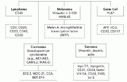
Diagram 45-1 Immunochemical approach to the classification of undifferentiated tumors. Closed boxes indicate recommended antibodies for primary screening of cases. Dashed boxes indicate antibodies for additional characterization of these tumors.

The cytokeratins are distributed in tissue specific patterns and both primary and metastatic tumors tend to recapitulate the cytokeratin profiles of the tissues from which they are derived (Wang et al, 1995). In some cases, patterns of cytokeratin expression are simple, while in others, complex patterns of cytokeratin expression are apparent. Hepatocellular carcinomas, for example, express cytokeratins 8 and 18 while multiple acidic and basic cytokeratins are found in squamous carcinomas. In some instances, specific cytokeratins can be used to differentiate cells and tumors of particular types. For example, Merkel cell tumors are typically positive for cytokeratin 20 (in addition to other cytokeratins) while small cell carcinomas arising from other sites are cytokeratin 20 negative (Moll et al, 1992). In some instances, the subcellular localization of cytokeratin immunoreactivity may provide important diagnostic clues. For example, small carcinomas often show a paranuclear dot-like staining pattern while mesotheliomas show a perinuclear net-like pattern of staining.

There is now an extensive literature on the distribution of cytokeratins in epithelial and non-epithelial tumors. Generally, low molecular weight cytokeratins are present in simple and glandular epithelium while high molecular weight cytokeratins are typical of stratified epithelium. Wang et al as well as other authors have studied the coordinate expression of CK7 (54kD) and CK20 (46kD) in a large series of carcinomas of diverse origins (Wang et al, 1995; Chu et al, 2000; Tot, 1999; Blumenfeld et al, 1999) (Diagram 45-2). CK7 is distributed in a wide array of normal simple epithelia while CK20 is present in normal intestinal epithelium, gastric foveolar cells, urothelial umbrella cells and Merkel cells (Figs. 45-1 and 45-2). Tumors that coexpress both CK7 and CK20 include urothelial (transitional cell) carcinomas of the bladder and pancreatic adenocarcinomas while CK7 and CK20 negative tumors include hepatocellular, prostatic and renal cell carcinomas as well as squamous and neuroendocrine lung carcinomas. The CK7/CK20+ phenotype is characteristic of adenocarcinomas of colorectal origin and Merkel cell tumors while the CK7+/CK20 phenotype is characteristic of tumors arising from a wide variety of other sites including the ovary, endometrium, breast and lung as well as mesotheliomas. Some carcinomas, including primary gastric adenocarcinomas typically show considerable heterogeneity in the expression patterns of cytokeratins 7 and 20.

Cytokeratin 14 is an acidic cytokeratin which is restricted in its distribution to the myoepithelial cells of a variety of organs and basal cells of kerantinized squamous epithelium (Chu et al, 2001). This marker is useful for the distinction of poorly differentiated squamous cell carcinoma from other poorly differentiated carcinomas. The studies of Chu et al have demonstrated that CK14 is present in most cases of squamous cell carcinoma (irrespective of their sites of origin or degrees of differentiation), neoplasms with focal squamous differentiation (endometrial and ovarian adenocarcinomas, mesotheliomas and transitional cell carcinomas), thymomas, myoepithelial components of salivary gland pleomorphic adenomas, and oncocytic tumors.

Coexpression of cytokeratins and other intermediate filaments is relatively common in carcinomas. For example, renal, endometrial and thyroid follicular neoplasms, as well as mesotheliomas, often coexpress cytokeratins and vimentin while medullary thyroid carcinomas, carcinoids, and pancreatic endocrine tumors may coexpress cytokeratins, vimentin and neurofilament proteins (Fig. 45-3). Small cell desmoplastic tumors often coexpress cytokeratins together with vimentin and desmin.

Cytokeratin expression has also been documented in a number of sarcomas including synovial and epithelioid sarcomas, leiomyosarcomas, rhabdomyosarcomas, chondrosarcomas
and Ewing’s tumor. Melanomas, particularly in metastatic sites, may also be positive for cytokeratins and occasional examples of malignant lymphoma, particularly of the Ki-1 and large cell types, have been reported to be positive for cytokeratins. Immunoreactivity for cytokeratins (AE1/AE3) also occurs in glioblastoma multiforme and this finding has been attributed to cross reactivity with epitopes present in the neoplastic glial cells (Morrison and Prayson, 2000). This is particularly problematic when the differential diagnosis includes metastatic poorly differentiated carcinoma. Oh and Prayson (1999) have shown that more than 50% of cases of glioblastoma multiforme are reactive for AE1/AE3 while only 1 of 23 cases contained cells that were positive for CAM5.2 and cytokeratins 7 and 20. In contrast, metastatic carcinomas were positive both for AE1/AE3 and CAM5.2.


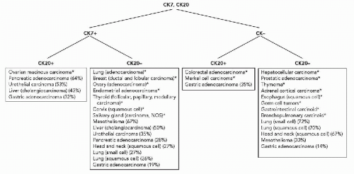
Diagram 45-2 Differential expression of cytokeratins 7 and 20. *More than 75% of cases are positive or negative. (Data from Wang et al, 1995; Blumenfeld et al, 1999; Tot, 1999; Chu et al, 2000A.)


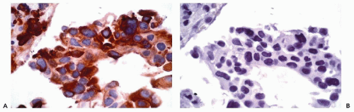
Figure 45-1 Poorly differentiated adenocarcinoma of the lung. A. Immunoperoxidase stain for cytokeratin 7 in cell block demonstrates uniform positivity within the tumor cells. B. Immunoperoxidase stain for cytokeratin 20 is negative.


Epithelial Membrane Antigen (EMA)

The milk fat globule membrane antigens represent a family of highly glycosylated proteins that are present on the apical membranes of breast epithelial cells. Antisera raised against these proteins include antibodies to the epithelial membrane antigen (EMA), a 70Kd glycosylated protein. EMA is not restricted in its distribution to breast epithelial cells but is present in a very wide variety of epithelial cells (Pinkus and Kurtin, 1985; Singh et al, 1995). In addition, a variety of normal and neoplastic lymphoreticular cells are reactive
for EMA. Perineural cells are also reactive for EMA and positive reactions for this marker are present in nerve sheath tumors. Other tumors that are positive for EMA include meningiomas and certain sarcomas (epithelioid sarcoma, synovial sarcoma, chordoma, and chondrosarcoma) (Fig. 45-4).


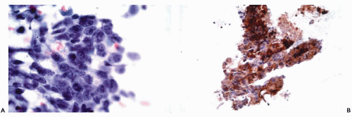
Figure 45-2 Metastatic colonic adenocarcinoma in liver. A. Fine needle aspiration biopsy stained with the Papanicolaou stain. B. Immunoperoxidase stain for cytokeratin 20 in cell block demonstrates uniform positivity within the tumor cells.


Carcinoembryonic Antigen

Carcinoembryonic antigen (CEA) is a highly glycosylated protein which was detected initially in fetal gut and colonic adenocarcinomas (Fig. 45-5). There are considerable variations in staining patterns with different polyclonal and monoclonal CEA antibodies (Sheahan et al, 1990). In part, these differences are related to the presence of antibodies which cross-react with nonspecific cross-reacting antigens (NCA) that are widely distributed in tissues. In immunohistochemical preparations utilizing polyclonal antisera or monoclonal antibodies, the presence of reactivity in granulocytes indicates the presence of NCAs; however, monoclonal antibodies may be selected which lack immunoreactivity for NCAs. CEA is present in a variety of normal cells and epithelial neoplasms while melanomas, lymphoma, and sarcomas are negative.


Other Epithelial Markers

A variety of other epithelial markers have been developed for use in immunocytochemistry, including B72.3, leu M1 (CD15), MOC-31, and BerEP4 which react with glycoprotein antigens (Diagram 45-3). The antigen recognized by B72.3 is a plasma membrane glycoprotein which is known as the tumor associated glycoprotein (TAG)-72. B72.3 immunoreactivity is present in a wide spectrum of carcinomas and some benign tissues (e.g., endometrium) but is absent in mesotheliomas, germ cell tumors and carcinomas of the adrenal cortex, liver, kidney, nasopharynxl, and thyroid (Thor et al, 1986; Loy et al, 1993). Antibodies directed to leu M1 (CD15) react with neutrophils, monocytes, a subset of T cells and Reed Sternberg cells of classic Hodgkin’s lymphoma (Arber and Weiss, 1993). In addition, CD15 immunoreactivity is present in approximately 75% of pulmonary adenocarcinomas and in a high proportion of carcinomas of the breast, kidney, and ovary, while mesotheliomas are typically negative (Sheibani et al, 1986). BerEP4 represents another glycoprotein antigen which is expressed in a wide variety of epithelial malignancies including those of the GI tract, pancreatico-biliary tract, breast, ovary, and pancreas while mesotheliomas are usually negative (Fig. 45-5) (Sheibani et al, 1991). MOC31, which is discussed in further detail in the section on mesothelioma, is also positive in a wide range of epithelial malignancies but is nonreactive with germ cell malignancies, hepatocellular carcinoma, renal cell carcinoma and mesothelioma (Ruitenbeek et al, 1994). The expression of these markers, however, is often focal and a negative result may be related to sampling, a problem exacerbated in cytological specimens.

Other epithelial markers which have been used in the diagnostic setting include CA125, CA19-9 and CA15-3. CA125 (OC125) was initially defined in ovarian carcinoma cell lines and is most commonly expressed in müllerian neoplasms, mesotheliomas and bile duct carcinomas, but may also be found in a small proportion of other tumor types, including those of the breast and thyroid (Bast et al, 1981; Kabawat et al, 1983; Haglund, 1986) (Fig. 45-6). CA19-9 is present in carcinomas of the gastrointestinal tract, pancreatico-biliary system, and müllerian origin; however, it is also expressed in a high proportion of other tumor types, including those of thyroid origin (Loy et al, 1993). CA15-3 is expressed in many tumor types, including those of müllerian, gastrointestinal, pancreaticobiliary, renal, breast, and prostatic origin (Gatalica and Miettinen, 1994).


Transcription Factors as Selective Epithelial Markers

Transcription factors are proteins that bind to regulatory elements in the promoter and enhancer regions of DNA
and stimulate or suppress gene expression. Transcription factors may be tissue specific or may be present in a variety of different tissue types. Thyroid transcription factor-1 (TTF1), for example, is present in thyroid follicular cells and C-cells and is also present in the lung (Ordonez, 2000) (Fig. 45-7). The adrenal 4 site/steroidogenic factor is present in steroid-producing cells and in certain anterior pituitary cell types. The pituitary transcription factor, Pit-1, is present in certain cells of the anterior pituitary and is also present in the placenta (Kulig and Lloyd, 1996). In some instances, antibodies to transcription factors are of considerable value in determining the origins of tumors of unknown primary sites.


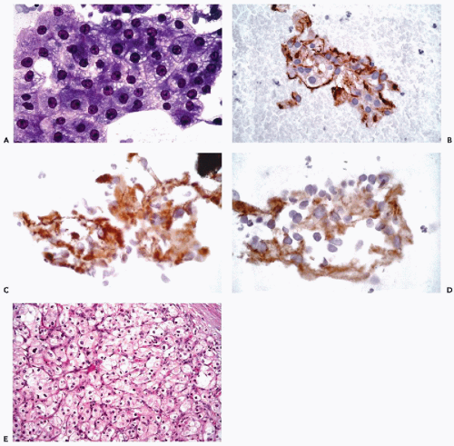
Figure 45-3 Metastatic renal cell carcinoma involving the lung. A. Fine needle aspiration biopsy stained with the Papanicolaou stain. B. Immunoperoxidase stain for EMA performed on destained slide demonstrates membrane positivity. C. Immunoperoxidase stain for cytokeratins 8 and 18 (CAM5.2) on a de-stained slide demonstrates cytoplasmic positivity. D. Immunoperoxidase stain for vimentin on a destained slide demonstrates focal cytoplasmic positivity. E. Histological section stained with hematoxylin and eosin demonstrates the typical features of renal cell carcinoma.


MESENCHYMAL MARKERS


Vimentin

Vimentin (MW57000) is the characteristic intermediate filament type of mesenchymal cells and their corresponding tumors (Battifora, 1991) (see Chap. 2, Diagram 45-2 and Fig. 45-4). The presence of immunoreactive vimentin in stromal and endothelial cells is considered an index of adequate tissue fixation and has been used as an internal “control” for this purpose. Vimentin is not restricted in its distribution to mesenchymal cells, however, since it is also found in a wide variety of normal and neoplastic epithelial and neural type cells. In some instance, mesenchymal cells and their tumors such as thyroid and kidney also contain cytokeratin proteins as discussed below (Miettinen, 1987; Suster, 2000). In mesenchymal cell neoplasms, immunochemical analysis of other markers in addition to vimentin provides evidence of specific lines of differentiation.


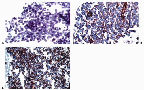
Figure 45-4 Metastatic synovial sarcoma involving the lung. A. Fine needle aspiration biopsy stained with the Papanicolaou stain. B. Immunoperoxidase stain for EMA in cell block demonstrate plasma membrane positivity. C. Immunoperoxidase stain for vimentin in cell block demonstrates diffuse cytoplasmic positivity.


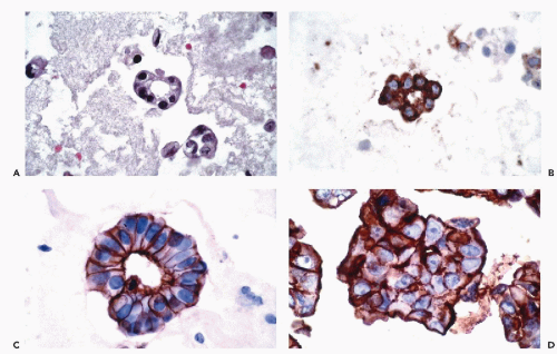
Figure 45-5 Metastatic adenocarcinoma in pleural effusion. A. Hematoxylin and eosin stain of cell block. B. Immunoperoxidase stain for CEA in cell block reveals cytoplasmic and membrane positivity. C,D. Immunoperoxidase stain for B72.3 also demonstrates plasma membrane positivity.


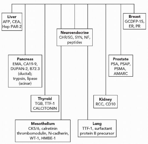
Diagram 45-3 Immunocytochemistry of poorly differentiated carcinomas. AFP, alpha fetoprotein; AMARC, alpha-methylacyl-coAracemase; CEA, carcinoembryonic antigen; CHR/SG, chromogranins/secretogranins; EMA, epithelial membrane antigen; ER, estrogen receptor; GCDFP-15, gross cystic disease fluid protein-15; HepPar 1, hepatocyte paratin antibody; NF, neurofilaments; PSA, prostate-specific antigen; PSMA, prostate-specific membrane antigen; PSAP, prostate-specific acid phosphatase; PR, progesterone receptor; RCC, renal cell carcinoma; SYN, synaptophysin; TGB, thyroglobulin; TTF1, thyroid transcription factor; WT-1, Wilms’ tumor gene protein.


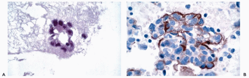
Figure 45-6 Ovarian papillary serous carcinoma involving the omentum. A. Hematoxylin and eosin stain of cell block. B. Immunoperoxidase stain for CA-125 in cell block demonstrates plasma membrane positivity.


Muscle Proteins

A variety of markers have been used to identify skeletal muscle differentiation including desmin, muscle-specific
actin (HHF35), skeletal muscle or sarcomeric actin, myoglobin, and creatine phosphokinase-MM (Tsukada et al, 1987; Swanson and Wick, 1995; Suster, 2000) (Fig. 45-8). Desmin (MW-55,000) is the major intermediate filament type of muscle cells including cardiac, skeletal and smooth muscle cell types. Both desmin and muscle-specific actin are the most commonly used markers for muscle tumors (Truong et al, 1990; Rangdaeng et al, 1991). Although myoglobin is specific for skeletal muscle, its sensitivity for the diagnosis of rhabdomyosarcoma is low, particularly in poorly differentiated forms of the tumor (Tsokos, 1994). An alternative approach for the identification of cells with skeletal muscle differentiation involves the use of myogenic regulatory proteins (Li and Olson, 1992). Myogenic regulatory proteins play a key role in the commitment of primitive mesenchymal cells to a skeletal muscle lineage. Since these proteins are expressed earlier than structural proteins such as actin, myosin and desmin, they are particularly useful for the diagnosis of rhabdomyosarcoma. Antibodies to myoD1 and myogenin have been used extensively for this purpose (Wang et al, 1995). In exceptional cases, rhabdomyosarcomas may be positive for cytokeratins, neurofilament triplet proteins, neuron-specific enolase, S100 protein and leu 7.


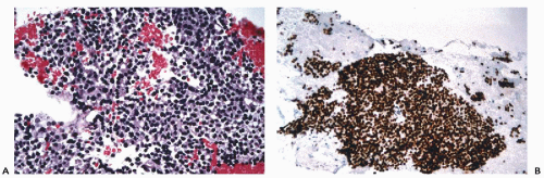
Figure 45-7 Metastatic poorly differentiated thyroid carcinoma in the lung. A. Cell block of fine needle aspiration biopsy stained with hematoxylin and eosin. B. Immunoperoxidase stain for thyroid transcription factor-1 in cell block demonstrates nuclear positivity.


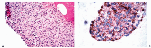
Figure 45-8 Metastatic leiomyosarcoma of the bladder involving the sacrum. A. Hematoxylin and eosin stain of cell block. B. Immunoperoxidase stain for smooth muscle actin in cell block demonstrates cytoplasmic positivity.

Tumors derived from smooth muscle are reactive with antibodies to desmin and muscle-specific actin (HHF35). Moreover, these tumors are also reactive with antibodies to smooth muscle actin and caldesmon (Watanabe et al, 1999). In some cases, smooth muscle tumors are also reactive with cytokeratin antibodies (Suster, 2000).


Vascular Markers

The factor VIII related antigen (von Willebrand’s factor) and Ulex europaeus I (UEAI) lectin have been used extensively for the evaluation of vascular tumors (Leader et al, 1986). Despite its high specificity, however, the factor VIII-related antigen has a relatively low sensitivity, particularly in poorly differentiated vascular tumors. UEAI, on the other hand, has high sensitivity but relatively low specificity since it reacts with a variety of epithelial cells. Although thrombomodulin was originally proposed as a marker for
tumors with endothelial differentiation, it is expressed in a variety of other tumor types (Ordonez, 1997). More recently, antibodies to CD31 and CD34 have been used for the identification of endothelial differentiation (Miettinen et al, 1994; von der Rijn and Rouse, 1994). It should be recognized, however, that a variety of soft tissue neoplasms, including dermatofibrosarcoma protuberans, solitary fibrous tumor, GI stromal tumors and peripheral nerve sheath tumors are also reactive for CD34. CD31, on the other hand, may be expressed weakly in some carcinomas and mesotheliomas (Suster, 2000).


Other Markers

Fibrohistiocytic tumors are vimentin positive and are variably reactive for alpha-1-antitrypsin, alph-1-antichymotrypsin and CD68 (Swanson and Wick, 1995). Occasional fibrohistiocytic tumors also exhibit immunoreactivity for cytokeratins. Neurogenic tumors, including Schwannomas, neurofibromas and malignant peripheral nerve sheath tumors are typically positive for S100 protein and are variably reactive for leu 7 (Wick et al, 1987; Johnson et al, 1988). EMA may also be present, particularly in plexiform neurofibromas. Both liposarcomas and chondrosarcomas are typically positive for S100 protein while osteogenic sarcomas are often positive for osteonectin. Sarcomas which consistently exhibit cytokeratin positivity include synovial and epithelioid sarcomas (Swanson and Wick, 1995; Suster, 2000). Chordomas are also typically positive for cytokeratins and EMA but also react with antibodies to S100 protein.

Bcl-2 expression was originally described in malignant lymphomas but was subsequently identified in a wide variety of other tumors including certain carcinomas and sarcomas (Joensun et al, 1994; Nakamura et al, 1997; Nakanishi et al, 1997). As noted by Suster (2000), the most useful applications of this antibody are in the diagnosis of the monomorphic variant of synovial sarcoma, solitary fibrous tumors, and gastrointestinal stromal tumors (GIST). Other connective tissue neoplasms that exhibit bcl-2 positivity include spindle cell lipoma, dendritic myxofibrolipoma Kaposi’s sarcoma, benign and malignant nerve sheath tumors, fibrosarcoma, low grade myxofibrosarcoma, malignant fibrous histiocytoma and dermatofibrosarcoma protuberans. CD99 is another marker which is expressed commonly in soft tissue tumors, including mesenchymal chondrosarcoma, synovial sarcoma, leiomyosarcoma, malignant fibrous histiocytoma and solitary fibrous tumor (Renshaw, 1995).

CD117 (C-kit) has emerged as an important marker for the interstitial cells of Cajal and the gastrointestinal stromal tumors which are thought to arise from these cells (Kindblom et al, 1998; Suster, 2000). However, c-kit expression also occurs in acute myeloid leukemia, mast cell disease, malignant melanoma, Ewing’s tumor, and a variety of carcinomas, including those of the breast, endometrium, lung, ovary and thyroid. Among spindle cell tumors, c-kit expression has been documented in leiomyosarcomas, dermatofibrosarcoma protuberans, hemangiopericytoma, malignant fibrous histiocytoma, and synovial sarcoma, among others.


LYMPHOID MARKERS

The malignant lymphomas include a heterogeneous group of neoplasms which have been classified into Hodgkin and non-Hodgkin types (see Chap. 31). The subclassification of these neoplasms depends on distinctive architectural, cytological, immunophenotypic and molecular features (Hughes et al, 1998; Jaffe-Perez et al, 1999). Immunophenotypic methods include flow cytometry and immunocytochemistry (see Chap. 47 and Diagram 45-2). The advantages of immunocytochemistry, particularly when applied to cytospin preparations, are the requirements for relatively small numbers of cells and the ability to directly correlate cellular morphology with patterns of marker expression (Simsir et al, 1999). Disadvantages of immunocytochemistry on cytospin preparations include the relatively low sensitivity of detection of small monoclonal populations and the inability to provide quantitative data. In institutions with considerable experience in immunocytochemistry and cytological specimens, the rate of correlation between flow cytometry and immunocytochemistry is as high as 98% (Simsir et al, 1999). In 98 cases reported by Simsir et al, 11% could not be phenotyped by flow cytometry and 4% could not be phenotyped by immunochemistry. While the precise subclassification of malignant lymphomas cannot be achieved in cytological preparations alone, their distinction from other types of poorly differentiated malignancies and reactive lymphoid proliferations can be accomplished in cytological samples.

One of the most useful markers for this purpose is leukocyte common antigen (CD45) (Chu et al, 2000) (Fig. 45-9; see Diagram 45-2). CD45 is a 200 kD glycoprotein which is present in the plasma membranes of B and T lymphocytes, monocytes and granulocytes but which is absent from erythrocytes and megakaryocytes. CD45 is present in the vast majority of malignant lymphomas although its reactivity may be weak or absent from plasmacytic neoplasms, anaplastic large cell lymphomas, Reed Sternberg cells in Hodgkin lymphomas of mixed cellularity, and nodular sclerosing and lymphocyte depleted Hodgkin lymphoma. Reed-Sternberg cells of lymphocyte-predominant Hodgkin lymphomas are usually positive. A specificity of 100% and a sensitivity of 90% have been reported for antibodies to CD45.

The immunophenotypic identification of B-cell lymphomas is accomplished by the demonstration of B-cell cell markers and in some cases (small lymphocytic lymphoma and mantle cell lymphoma) by the concurrent expression of CD5. The cases which are most amenable to cytological diagnosis are those that consist of a monomorphic cell population that does not require architectural assessment. Cases of follicular lymphoma, marginal zone lymphoma, mantle zone lymphoma, and Hodgkin lymphoma often require histological
assessment (see Chap. 31). B-cell lineage is most effectively accomplished by the application of CD19, CD20, CD22, and CD45RA antibodies (Fig. 45-9). In addition to these markers, the diagnosis of B-cell lymphomas and their differentiation from reactive processes requires the demonstration of light chain restriction by staining for kappa and lambda light chains. Monoclonality is defined as a kappa:lambda ratio of more than 6:1 or a lambda:kappa ratio of more than 4:1 (Sneige, 1990). In addition to CD20, antibodies that are of particular value in the categorization of mature B-cell lymphomas include CD43, CD5, CD10, CD23, and cyclin D1. Positive staining for CD43 and CD5 is characteristic of small lymphocytic lymphoma and mantle cell lymphoma. CD23 is consistently expressed in small lymphocytic lymphomas but is present in less than 20% of mantle cell lymphomas and marginal zone lymphomas. Among lymphomas, CD10 expression is limited to those of follicular type while cyclin D1 positivity is characteristic of mantle cell lymphomas.


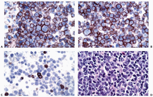
Figure 45-9 Large cell lymphoma. A. Immunoperoxidase stain for leukocytes common antigen (CD45) performed on a Papanicolaou stained aspirate demonstrates positive staining of the plasma membranes. B. Immunoperoxidase stain for CD20 demonstrates a similar pattern of staining. C. Immunoperoxidase stain for CD3 demonstrates a population of non-neoplastic T-cells. D. Hematoxylin and eosin stained section of a subsequent excisional biopsy demonstrates the typical features of a large cell lymphoma.

The diagnosis of T-cell lymphomas is considerably more difficult than the diagnosis of B-cell lymphomas in cytological preparations. The identification of T-cell lymphomas requires the demonstration of appropriate T-cell markers with dropout of certain of the antigens. The presence of terminal transferase is characteristic of lymphoblastic lymphomas. For further extensive discussion of these issues, see Chapter 31.


NEURAL AND NEUROENDOCRINE MARKERS


Neurofilaments

The neurofilaments are composed of heteropolymers of three different subunits with molecular weights of 70, 170, and 200 kD, corresponding to low (L), medium (M), and high (H) molecular weight subunits (Shaw and Weber, 1982; Kimura et al, 1990; Morrison and Prayson, 2000) (see Diagram 45-3). Each isoform differs by the extent of phosphorylation. The neurofilaments represent the major intermediate filaments of mature and developing neurons, paraganglionic cells and certain normal neuroendocrine cells. In neurons, NF-L and NF-M are present in immature cells with neuronal differentiation while NF-H is present in mature neurons. These proteins are also expressed in tumors with evidence of neuronal differentiation and are present in varying degrees in neuroendocrine tumors of epithelial type which also contain cytokeratins. Neuroendocrine
tumors of nonepithelial type, such as paragangliomas and neuroblastomas, are positive for neurofilaments but negative for cytokeratins (Wick, 2000; DeLellis, 2001).


Cytosolic Markers

Neuron-specific enolase (NSE) is a glycolytic dimeric enzyme composed of alpha, beta and gamma subunits. Neurons and neuroendocrine cells contain the gamma-gamma form of the enzyme while other forms of enolase are present in a wide variety of normal and neoplastic cells (Tapia et al, 1981). Since NSE is a cytosolic marker, it is usually positive even in those cells and tumors that contain few or no secretory granules. Although most antibodies to neuron specific enolase provide a high level of sensitivity for the detection of cells with neural/neuroendocrine differentiation, their specificity is low (Fig. 45-10) (Haimoto et al, 1985). Monoclonal antibodies to NSE, on the other hand, exhibit a higher specificity but lower sensitivity (Thomas et al, 1987). Protein gene product 9.5 (PGP 9.5) is another cytosolic marker that separates ubiquitin from other proteins (Rode, 1983). This marker is present in a wide variety of neuroendocrine cells and tumors but its specificity as an immunochemical marker is low.


Chromogranins

The chromogranins include a family of proteins which form the most abundant constituent by weight of the chromaffin granules (Lloyd and Wilson, 1983) (Fig. 45-11). The chromogranin family includes chromogranin A, chromogranin B, and secretogranin II, in addition to other chromogranin types (secretogranin IV and V) (Fahrenkamp et al, 1995). There is differential expression of chromogranins in normal cells and corresponding neoplasms of differing origins. For example, chromogranin A predominates in mid-gut carcinoids while chromogranin B is the predominant granin of hindgut neuroendocrine cells and carcinoids. The extent of chromogranin positivity generally parallels the numbers of intracytoplasmic secretory granules.


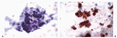
Figure 45-10 Small cell bronchogenic carcinoma. A. ThinPrep stained with Papanicolaou stain. B. Immunoperoxidase stain for neuron specific enolase on ThinPrep. The tumor cells were also positive for EMA and synaptophysin.


Synaptophysin

Synaptophysin is a 38 kD protein present in synaptic vesicles of neurons, neuroendocrine cells and many neuroendocrine tumors (Gould et al, 1986) (see Fig. 45-11). Synaptophysin, however, is not entirely specific for neuroendocrine tumors. Adrenal cortical neoplasms, for example, may exhibit synaptophysin reactivity (Komminoth et al, 1995).


Peptide Hormones and Amines

The hormonal content of neuroendocrine cells and tumors can be demonstrated effectively with monoclonal antibodies using a variety of staining formats (DeLellis, 2001). Although tumors of specific sites have characteristic patterns of hormone expression (see Chap. 39 as an example), the presence of a particular hormone does not allow absolute predication of the site of origin of a metastatic neoplasm. Calcitonin, for example, is present in medullary thyroid carcinomas, small cell lung carcinomas, bronchopulmonary carcinoids, and thymic and prostatic neuroendocrine tumors. Somatostatin shows a similar widespread distribution.


Other Markers

Leu 7 (CD57) reacts with lymphocytes with natural killer and killer cell activity, some neural and neuroendocrine cells and a wide variety of neoplasms including small cell carcinomas, neuroendocrine tumors, and carcinomas of the thyroid and prostate (Arber and Weiss, 1995).


GLIAL MARKERS

Glial fibrillary acidic protein (GFAP) (52 kD) is the major intermediate filament of fibrous and protoplasmic astrocytes and their corresponding neoplasms (Eng et al, 1971). This protein is also present in some ependymal cells, cerebellar radial glial cells and Müller cells of the retina.
While mature oligodendrocytes do not contain GFAP, cells that transiently express GFAP and myelin basic protein have been described. Staining intensity in glial tumors is inversely proportional to tumor grade (Schiffer et al, 1986; Morrison and Prayson, 2000). GFAP is also present in nonmyelinated Schwann cells, certain cells of the pituitary and breast and in tumors not considered to be of glial origin including mixed tumors of salivary gland and skin origin, nerve sheath tumors and chordomas.


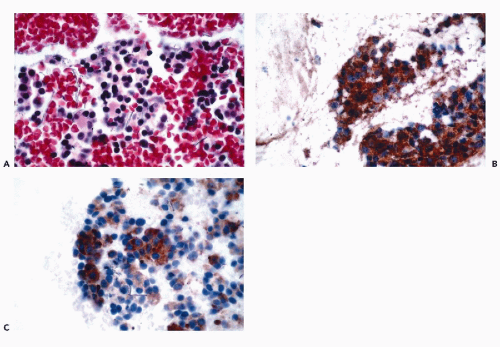
Figure 45-11 Pancreatic endocrine tumor. Cell block of fine needle aspirate stained (A) with hematoxylin and eosin, (B) with immunoperoxidase stain for chromogranin, and (C) with synaptophysin both showing cytoplasmic granularity.

S100 protein (MW20-25 kD) is a calcium binding protein composed of alpha and beta subunits (Jensen et al, 1985). The exact function of this protein is, however, unknown. S100 protein is present in a wide variety of cells in the nervous system including astrocytes, oligodendrocytes, ependymal cells, Schwann cells, and some neurons and in their corresponding neoplasms. At the cellular level, it is present both in the nuclei and in cytoplasm. This antigen is present in a wide variety of other normal cell types, including Langhans cells, and it is present in a very wide variety of tumor types including melanomas and carcinomas (Nakajima et al, 1982).


MELANOMA MARKERS

The diagnosis of malignant melanoma, particularly those tumors of amelanotic type, is challenging both in histological and cytological materials (Gupta and Lallu, 1997) (see Diagram 45-1). Numerous antibodies have been assessed for the diagnosis of this tumor, but none is absolutely specific. In most studies, a panel of antibodies directed to vimentin, S100 protein, HMB45, and MART1 (melanoma-associated antigen recognized by T cells) is used to establish the diagnosis of melanoma (Fig. 45-12; see Diagram 45-2).

Vimentin is strongly expressed in melanomas, but by itself this marker is of little diagnostic value since it is expressed in a wide variety of tumors (Angeli et al, 1988). Although initial studies indicated that melanomas were negative for cytokeratins, more recent studies employing histological and cytological materials have revealed cytokeratin positivity in variable numbers of cases, particularly in metastatic sites. In fine needle aspirates, for example, cytokeratin positivity has been found in up to 25% of evaluable cases (Banks et al, 1995).

S100 protein is the most sensitive marker for the diagnosis of melanoma; however, this protein is widely expressed in a variety of normal cells and neoplasms of diverse origins. Both in cytological and histological samples, positive staining is present, both within the nucleus and the cytoplasm. However, the value of S100 as a melanoma marker appears to be somewhat limited in alcohol-fixed FNA specimens (Simmons and Martin, 1991).

HM45 has a higher level of specificity but lower sensitivity than S100 (Simmons and Martin, 1991). The staining is typically granular and is present within the cytoplasm. Desmoplastic and spindle cell melanomas are usually negative for HMB45. Other lesions that are reactive for HMB45
include angiomyolipoma and pulmonary lymphangioleiomyomatosis (Zamecnik, 1999).


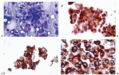
Figure 45-12 Metastatic malignant melanoma involving the lung. A. Diff-Quik stain. B. Immunoperoxidase stain for S100 protein demonstrates nuclear and cytoplasmic positivity. C. Immunoperoxidase stain for Melan-A (A103) demonstrates cytoplasmic positivity. D. Immunoperoxidase stain for HMB45 demonstrates cytoplasmic positivity.

Jun 8, 2016 | Posted by in PATHOLOGY & LABORATORY MEDICINE | Comments Off on Immunochemistry and Molecular Biology in Cytological Diagnosis

Full access? Get Clinical Tree

Get Clinical Tree app for offline access