Low-magnification view of a hobnail hemangioma (HH) demonstrates superficial dilated vascular spaces  in the papillary dermis with deeper small blood vessels and stromal hemosiderin deposits
  .
.
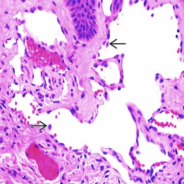
Higher power examination of the superficial portion of an HH shows superficial dilated vessels lined by small endothelial cells with nuclear hyperchromasia protruding into the lumina
 .
.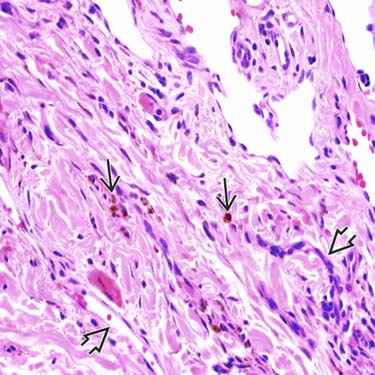
Histologic examination of the deeper aspect of the lesion shows small, thin-walled blood vessels
 and prominent hemosiderin deposition in the stroma
  .
.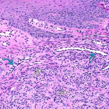
This is an example of a more cellular HH composed of many closely apposed vessels in the superficial dermis, many of which show very small, compressed lumens
 . Hobnailed endothelial cells can be appreciated in some of the vessels
  .
.
CLINICAL ISSUES
Epidemiology


Presentation
• Skin papule or nodule


Stay updated, free articles. Join our Telegram channel
Full access? Get Clinical Tree
