Patient Story
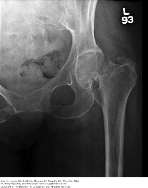
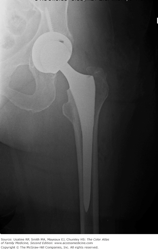
A 60-year-old woman comes to the emergency room for hip pain. She felt a pop in her hip accompanied by the immediate onset of pain that prohibited her from walking. She had fallen 2 days prior. Figure 105-1 shows a transcervical left femoral neck fracture with varus angulation and superior offset of the distal fracture fragment. She was evaluated by an orthopedic surgeon and underwent surgery the next day (Figure 105-2). After many months of rehabilitation, she was able to walk again.
Epidemiology
Risk Factors
Diagnosis
- In a population study, major risk factors for hip fracture include:
- Low bone mineral density (3.6-fold [95% confidence interval (CI), 2.6 to 4.5] in women and 3.4-fold [95% CI, 2.5 to 4.6] in men for each standard deviation [SD] [0.12 g/cm2] reduction in bone mineral density).5
- Postural instability and/or quadriceps weakness.
- A history of falls.
- Prior hip fracture.5
- Other factors associated with increased risk include: dementia, tobacco use, physical inactivity, impaired vision, and alcohol use.
- Low bone mineral density (3.6-fold [95% confidence interval (CI), 2.6 to 4.5] in women and 3.4-fold [95% CI, 2.5 to 4.6] in men for each standard deviation [SD] [0.12 g/cm2] reduction in bone mineral density).5
Physical examination—Abducted and externally rotated hip; limp or refusal to walk.
Typical Distribution
- Hip fractures are classified according to anatomic location.6
- Intracapsular (femoral neck fracture; Figures 105-1 and 105-3).
- Extracapsular (intertrochanteric or subtrochanteric fracture; Figure 105-4).
- Intracapsular (femoral neck fracture; Figures 105-1 and 105-3).


