• Classically, does not connect to epidermis and is deep-seated
• Solid areas composed of varying proportion of
Clear cells
Poroid cells
Squamoid cells
Rarely mucinous cells
• Ducts with eosinophilic cuticles present in solid areas
• Cystic areas lined by cuboidal cells
Sometimes with evidence of decapitation secretion
• Stroma between solid islands &/or cystic areas is prominently hyalinized
• Well circumscribed
• Necrosis usually absent
• Prominent cytologic atypia not present
• Mitoses not numerous
Top Differential Diagnoses
• Metastatic renal cell carcinoma
• Other clear cell tumors
Basal cell carcinoma
Squamous cell carcinoma
• Other adnexal tumors
Lymphadenoma
Sebaceous adenoma
Cystadenoma
Sebaceoma
• Hidradenocarcinoma
Hidradenoma at Low Magnification Low-magnification view of a hidradenoma shows a dermal-based, nodular-appearing, solid to cystic tumor.
Hidradenoma: Characteristic Stroma, Cells, and Ducts Hidradenoma often has a prominent hyalinized stroma . Islands are composed of clear cells and poroid cells with some evidence of ductal and glandular differentiation.
Hidradenoma: Poroid and Clear Cells This solid area of a hidradenoma is composed of small, bland-appearing clear cells and poroid cells . There is minimal cytologic atypia, an absence of necrosis, and mitoses are not prominent.
Hidradenoma: Poroid and Clear Cells Higher magnification of hidradenoma shows an admixture of clear cells and basaloid/poroid cells . The basaloid cells have minimal cytoplasm, which is eosinophilic to basophilic staining and round/oval nuclei. Mitoses and cytologic atypia are not present.


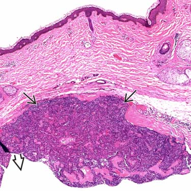
 to cystic
  tumor.
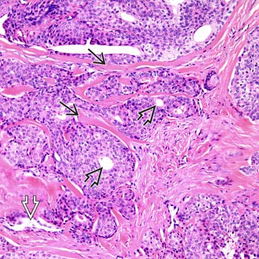
 . Islands are composed of clear cells and poroid cells with some evidence of ductal
  and glandular
  differentiation.
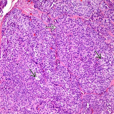
 and poroid cells
  . There is minimal cytologic atypia, an absence of necrosis, and mitoses are not prominent.
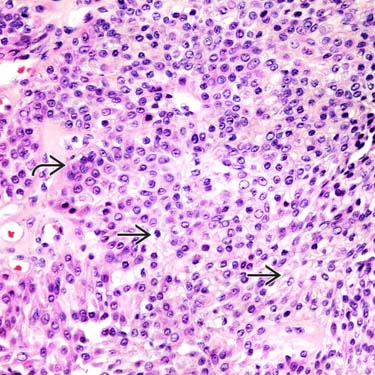
 and basaloid/poroid cells
  . The basaloid cells have minimal cytoplasm, which is eosinophilic to basophilic staining and round/oval nuclei. Mitoses and cytologic atypia are not present.


