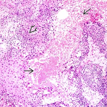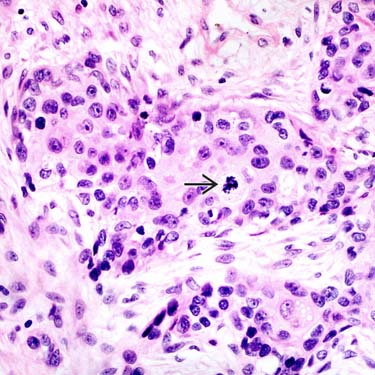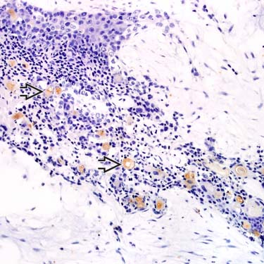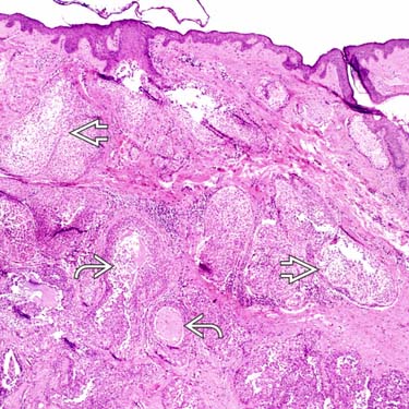
Local recurrences Metastases Microscopic • Epithelial islands with interspersed glands/ducts • Cells composing islands are variable Clear Poroid Squamoid (less common) Rarely mucinous • Hyalinized stroma Similar to hidradenoma • Infiltrative growth pattern Not well circumscribed, unlike hidradenoma • Deep extension • Perineural or vascular invasion may be present • Ki-67 generally shows high proliferative index Top Differential Diagnoses • Hidradenoma • Porocarcinoma • Metastatic renal cell carcinoma • Clear cell squamous cell carcinoma • Clear cell basal cell carcinoma • Sebaceous carcinoma Hidradenocarcinoma: Low MagnificationLow magnification of a hidradenocarcinoma shows a dermal-based, atypical, multilobular neoplasm with clear cell features and cystic spaces containing mucinous material and cellular debris. Hidradenocarcinoma: Areas of NecrosisAreas of squamous differentiation and prominent tumoral cell necrosis are present in this example of hidradenocarcinoma. Squamous differentiation is much less common than in porocarcinoma. Hidradenocarcinoma: Atypia and MitosesInfiltrative islands of tumor cells in hidradenocarcinoma show cellular atypia, with enlarged nuclei and prominent nucleoli. There is also an atypical mitosis present. Hidradenocarcinoma: CEA PositivityCEA highlights numerous small ductal lumina and their contents in this example of an infiltrative hidradenocarcinoma. TERMINOLOGY Synonyms • Malignant acrospiroma, malignant hidradenoma Definitions • Malignant adnexal tumor arising from, or showing areas similar to, hidradenoma CLINICAL ISSUES Site • Most frequently in head and neck region Presentation • Nodule or mass Treatment • Surgical approaches Wide, local excision of tumor Mohs micrographic surgery may be best option to ensure clear margins • Drugs For metastatic hidradenocarcinoma, if surgery is not option – Various chemotherapeutic regimens reported – Case reports of sunitinib and capecitabine treatment • Radiation Variable response Prognosis • Excision may be curative • Course can be aggressive Local recurrences, metastatic disease (to lymph nodes, distant skin, internal organs) MICROSCOPIC Histologic Features • Epithelial islands with interspersed glands/ducts • Architecture may be predominantly solid islands; sometimes cystic areas present Cells composing islands are variable – Clear, poroid, less commonly squamoid; rarely mucinous • Hyalinized stroma • Infiltrative growth pattern; not well-circumscribed • Deep extension • Necrosis may be evident • Nuclear pleomorphism may be present • Mitoses often seen May see few to many Only gold members can continue reading. Log In or Register to continue Share this:Click to share on Twitter (Opens in new window)Click to share on Facebook (Opens in new window)Like this:Like Loading... Related Related posts: Squamous Cell Carcinoma In Situ (Bowen Disease) Porocarcinoma Multinucleate Cell Angiohistiocytoma Glomus Tumors Stay updated, free articles. Join our Telegram channel Join Tags: Diagnostic Pathology: Neoplastic Dermatopathology Apr 24, 2017 | Posted by admin in PATHOLOGY & LABORATORY MEDICINE | Comments Off on Hidradenocarcinoma Full access? Get Clinical Tree