Hepatocellular Carcinoma in Cirrhotic Liver Autopsy specimen shows a large, central mass with small satellite tumor nodules . The latter may represent intrahepatic spread due to vascular invasion or independent primaries. The nonneoplastic liver shows cirrhosis.
Hepatocellular Carcinoma in Noncirrhotic Liver This image shows a unifocal, yellow-tan, well-circumscribed tumor in the background of a normal liver. Of hepatocellular carcinoma (HCC), 10-30% arise in noncirrhotic liver.
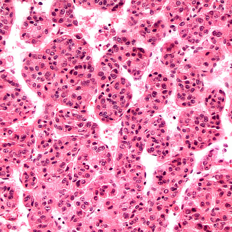
Trabecular Pattern Neoplastic cells resemble hepatocytes and have a high nuclear:cytoplasmic ratio. The tumor cells are organized in thick, disordered trabeculae.
Pseudoacinar Pattern Pseudoacinar or pseudoglandular pattern is common in HCC and can mimic adenocarcinoma. Unlike true glands, there is no basement membrane, and the nuclei do not have a basal location.
TERMINOLOGY
Abbreviations
• Hepatocellular carcinoma (HCC)
Synonyms
• Hepatoma
Definitions
• Primary malignant neoplasm of liver with hepatocytic differentiation
ETIOLOGY/PATHOGENESIS
Developmental Anomaly
• HCC can occur in patients with various congenital anomalies, including Alagille syndrome, ataxia-telangiectasia, Abernethy malformation, and genetic diseases such as bile salt export protein (BSEP) deficiency
Environmental Exposure
• Aflatoxin B1 (mycotoxin produced by fungi of Aspergillus genus that contaminates food) is major cause of HCC in China and southern Africa
• Alcoholic cirrhosis is major cause of HCC in western populations
• Other exposures linked to HCC include anabolic steroids, Thorotrast, oral contraceptives, and smoking
Infectious Agents
• Chronic viral hepatitis (hepatitis B and hepatitis C) is leading cause of HCC worldwide
Metabolic Disorders
• Various metabolic disorders, including hemochromatosis, tyrosinemia, hypercitrullinemia, α-1-antitrypsin deficiency, and fructosemia, are associated with increased risk of HCC
• Recent studies have implicated diabetes, obesity, and metabolic syndrome as risk factors
Cirrhosis
• 70-90% of HCC arises in cirrhosis
• Prognosis is significantly worse compared to HCC in noncirrhotic liver
• Macronodular cirrhosis is more strongly associated with HCC than micronodular
Progression of Benign Tumor
• HCC can arise in preexisting hepatocellular adenoma
CLINICAL ISSUES
Epidemiology
• Incidence
Varies widely depending on geography in parallel with prevalence of hepatitis B and C and aflatoxin exposure
– East Asia and southern Africa have highest incidence worldwide, up to 150 per 100,000
– In USA, annual incidence is ∼ 4 per 100,000
• Age
Incidence increases with advancing age and then falls off in elderly; however, average age varies depending on geography
– In parts of world with high incidence, average age is 35 years
– In USA, average age is 60 years
Can occur in children, particularly in those with metabolic or genetic disorders
• Sex
More common in men
Presentation
• Abdominal pain due to stretching of Glisson capsule
• Malaise, weight loss, hepatomegaly
• Decompensation of previously stable cirrhotic patient with jaundice and rapidly accumulating ascites
• Fever, leukocytosis, and liver mass mimicking hepatic abscess
• Increasingly, small asymptomatic tumors are being found during surveillance of cirrhotic patients
Laboratory Tests
• α-fetoprotein (AFP) is elevated in 70-90% of patients
Natural History
• Metastasis occurs in 40-60% of patients
Most common locations are lymph nodes in porta hepatis, around pancreas, and celiac axis
• HCC has tendency for intravascular spread with involvement of hepatic and portal veins
Hematogenous spread most commonly occurs to lungs, but also adrenal glands, bone, stomach, heart, pancreas, kidney, spleen, and ovary
• Tumor seldom breaches Glisson capsule, and, therefore, dissemination throughout peritoneal cavity is rare
Treatment
• Surgical approaches
Resection is possible if sufficient reserve liver function
Transplantation is option if patient meets Milan criteria of single tumor < 5 cm, or < 4 tumors, none > 3 cm
Less stringent UCSF criteria have been proposed: Solitary tumor < 6.5 cm, or < 4 tumors, none > 4.5 cm and total tumor diameter up to 8 cm, without gross vascular invasion
Histologic differentiation as selection criterion has been implemented in certain centers, as poor differentiation has been shown to be associated with high recurrence
• Drugs
HCC is resistant to chemotherapeutic agents
Sorafenib
– Tyrosine kinase inhibitor that has proven to be at least somewhat effective in advanced cases
• Ablation therapy
Radiofrequency or microwave ablation, or direct percutaneous ethanol injections are options for small tumors
Transarterial embolization (TEA) and transarterial chemoembolization (TACE) can prolong survival
Prognosis
• Favorable prognostic factors
Age < 50 years, female gender
Resectable tumor
Noncirrhotic liver
Encapsulated tumor, early HCC
Well or moderately differentiated
Absence of vascular invasion
• In USA, 5-year survival is 30% for localized disease, 10% for regional disease, and < 5% for metastatic disease
• For early cancers that receive transplant, 5-year survival is 60-70%
IMAGING
Radiographic Findings
• Characteristic features on contrast-enhanced study (dynamic CT scan or MR)
HCC enhances more intensely than surrounding liver in arterial phase
HCC enhances < surrounding liver in venous phase (washout)
• Biopsy not required for diagnosis if findings typical of HCC are seen in cirrhotic liver of lesions > 2 cm
• For lesions 1-2 cm, typical radiology findings on 2 techniques increases sensitivity and specificity of diagnosis
• All suspicious lesions in noncirrhotic liver as well as ones in cirrhotic liver with atypical imaging features should be biopsied
• Liver Imaging Reporting and Data System (LI-RADS) is now being used
Combines arterial enhancement with size, venous washout, presence of capsule and growth compared to prior imaging to yield 5 diagnostic categories
– LR-1: Definitely benign
– LR-2: Probably benign
– LR-3: Moderate probability of benign or malignant
– LR-4: Probably malignant
– LR-5: Definitely malignant
MACROSCOPIC
General Features
• Variable hemorrhage and necrosis, can be bile-stained
• Solitary ± satellite nodules, or multiple discrete tumors
• Multiple small, indistinct tumor nodules can mimic cirrhosis on imaging and gross examination (cirrhosis-like variant)
Pedunculated tumors are rare, more easily resected and have better prognosis
Encapsulated tumors are usually solitary tumors that arise in cirrhotic livers and have better prognosis
• Gross venous or bile duct invasion may be seen
MICROSCOPIC
Histologic Features
• Architectural patterns
Trabecular pattern: Tumor cells grow as thickened hepatic plates separated by sinusoids without desmoplastic stroma
Pseudoglandular or acinar pattern: Tumor cells grow in solid nests with central degenerative changes
Compact pattern: Trabeculae grow compressed together
Spindle cell pattern: Often referred to as sarcomatoid HCC
• Tumor cell morphology
Tumor cells resemble hepatocytes with polygonal shape, round vesicular nuclei, and prominent nucleoli
Inclusions can be seen in tumor cells: Mallory hyaline, ground-glass inclusions, hyaline globules, pale bodies
Clear cells may be present and even numerous due to accumulation of glycogen or fat
 . The latter may represent intrahepatic spread due to vascular invasion or independent primaries. The nonneoplastic liver shows cirrhosis.
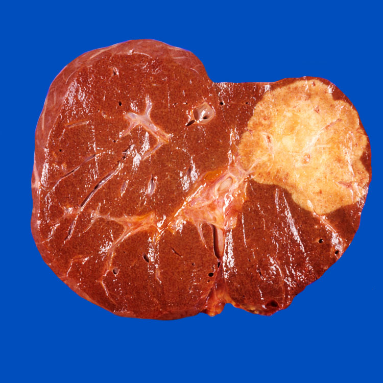

 Incidence increases with advancing age and then falls off in elderly; however, average age varies depending on geography


 Transplantation is option if patient meets Milan criteria of single tumor < 5 cm, or < 4 tumors, none > 3 cm
 Less stringent UCSF criteria have been proposed: Solitary tumor < 6.5 cm, or < 4 tumors, none > 4.5 cm and total tumor diameter up to 8 cm, without gross vascular invasion


 Trabecular pattern: Tumor cells grow as thickened hepatic plates separated by sinusoids without desmoplastic stroma
 Pseudoglandular or acinar pattern: Tumor cells grow in solid nests with central degenerative changes


