A number of landmarks visible on the body’s surface correspond to deeper structures. • Lies at level of C3 vertebra • Does not articulate with any other bone • Is suspended by muscles from • Bifurcation of common carotid artery into external and internal carotid arteries • Site of carotid sinus (baroreceptor) and carotid body (chemoreceptor) • Carotid pulse can be palpated at anterior border sternocleidomastoid (level of C5 vertebra). • Junction of larynx and trachea • Junction of pharynx and esophagus • Level at which inferior and middle thyroid arteries enter the thyroid gland • Vertebral artery (first branch subclavian artery) enters foramen transversarium of C6 transverse process to ascend to brain through successively higher foramina. • Superior belly of omohyoid muscle crosses carotid sheath. • Level of middle cervical sympathetic ganglion • Carotid artery can be compressed and palpated against transverse process C6. • Isthmus of thyroid gland overlies second and third tracheal cartilages. • Jugular (suprasternal) notch • Thin, broad sheet of muscle within superficial fascia of the neck • Muscle of facial expression, tensing the skin • Draws corners of mouth down, as in a grimace, and depresses mandible • Deep to platysma, descends from angle to mandible to midpoint of clavicle • Useful for assessment of venous filling with patient sitting at 45 degrees • Divides neck into anterior and posterior triangles (see Section 1.4, Head and Neck—Neck) • Sternal head attaches to manubrium of sternum. • Clavicular head attaches to superior middle third of clavicle. • Can be seen and palpated when acting unilaterally to flex and rotate head and neck to one side, so that ear approaches shoulder and chin turns in the opposite direction • Smooth midline prominence on the frontal bone • Located above the root of the nose, between supra-orbital margins • Inion—prominent point of external occipital protuberance at back of head • Auricle—part of external ear • Skeleton mainly cartilaginous • Dorsum extends from root to apex • Inferior surface has two openings or nares (nostrils) • Philtrum—midline infranasal depression of upper lip • Felt over ramus of mandible when teeth are clenched • Parotid duct can be palpated at medial border (duct opens over second molar inside cheek). • Temporalis muscle can be felt above zygomatic arch when teeth clenched. • Facial artery can be palpated over lower margin body of mandible in line with a point one fingerbreadth lateral to the angle of the mouth. • Transverse incision through skin of neck and anterior wall of trachea • Method for achieving a definitive airway • Transverse incision made through skin, at midpoint between suprasternal notch and thyroid cartilage • Platysma and pretracheal fascia divided • Thyroid isthmus divided or retracted • Opening made between first and second tracheal rings or through second through fourth tracheal rings • Large veins such as the subclavian have relatively constant relationships to easily identifiable anatomical landmarks. • Placement of large-bore venous catheter in an emergent situation to deliver high flow of fluid or blood products • Used for administration of chemotherapeutic agents, hyperalimentation fluids, and so on • Used for assessing right heart (venous) pressures • Vein located in an area bounded by the sternal and clavicular attachments of sternocleidomastoid and the clavicle—just deep to middle third of clavicle • Subclavian vein is inferior and anterior to subclavian artery and separated from it by anterior scalene muscle. • Encloses, supports, and protects brain and meninges • Contains foramina for transmission of nerves and vessels • Contains specialized cavities and openings for sense organs (e.g., nasal, oral) • Cranial vault and base of skull • Bones united by interlocking sutures • Calvaria composed of 4 bones • Encloses orbits, nose, paranasal sinuses, mouth, and pharynx • Maxillae and mandible form upper and lower jaw, respectively, and house the teeth • Coronal suture separates frontal and parietal bones. • Sagittal suture separates two parietal bones. • Lambdoid suture separates parietal and temporal bones from occipital bones. • Squamous suture separates squamous part of temporal bone from parietal bone. • Sphenosquamous suture separates squamous part of temporal bone from greater wing of sphenoidal bone. • Metopic suture between two frontal bones is largely obliterated with fusion of frontal bones • Divided into anterior, middle, and posterior cranial fossae • Contains frontal lobe of brain • Formed by frontal bone anteriorly, ethmoidal bone medially, and lesser wing of sphenoidal bone posteriorly • Frontal crest—midline bony extension of frontal bone • Foramen cecum—foramen at base of frontal crest • Crista galli—midline ridge of bone from ethmoid posterior to foramen cecum • Cribriform plate—thin, sievelike plate of bone on either side of crista galli, which transmits olfactory nerves from nasal cavity to olfactory bulbs • Contains temporal lobe, hypothalamus, and pituitary gland • Formed by greater wing and body of sphenoidal bone, petrous temporal bone, lesser wing of sphenoidal bone • Sella turcica—central depression in body of sphenoidal bone for pituitary gland • Tuberculum sellae—swelling anterior to sella turcica • Dorsum sellae—crest on body of sphenoidal bone posterior to sella turcica • Anterior clinoid processes—medial projections of lesser wings of sphenoidal bones • Posterior clinoid processes—swelling at either end of dorsum sellae • Foramen lacerum (one on each side)—jagged opening closed by plate of cartilage in life, transmits nothing • Contains four foramina in a crescent on either side in the body of the sphenoidal bone • Contains cerebellum, pons, and medulla oblongata • Composed largely of occipital bone, body of sphenoidal bone, petrous, and mastoid parts of temporal bone • Foramen magnum—transmits spinal cord • Internal occipital crest—divides posterior fossa into two lateral cerebellar fossae • Grooves for transverse and sigmoid dural venous sinuses • Jugular foramen—transmits sigmoid sinus (internal jugular vein) and several cranial nerves • Internal acoustic meatus—anterior and superior to jugular foramen, transmits facial and vestibulocochlear nerves (CN VII and CN VIII) • Hypoglossal canal—anterolateral and superior to foramen magnum, transmits hypoglossal nerve (CN XII) • Largest and strongest bone in face • Articulates with temporal bone at temporomandibular joint • Can be divided into lower base and upper alveolar part • Has mental protuberance anteriorly and inferiorly where two sides come together • Mental spine: rough projection on inner surface of body in midline • Mental foramen below second premolar transmits terminal branch of inferior alveolar nerve to supply skin and mucus membrane of lower lip and chin. • Mylohyoid line: ridge extending upward and backward on internal surface of alveolar part of mandible for attachment mylohyoid muscle • Submandibular fossa: long depression below mylohyoid line, which accommodates submandibular gland • Sublingual fossa: concavities on either side of mental spine for sublingual gland • Lateral vertical projections from body • Each meets body inferiorly at angle of jaw. • Two processes at superior end: coronoid process and condylar process • Coronoid process—attachment of temporalis muscle • Condylar process—part of temporomandibular joint • Mandibular notch—concavity between condylar and coronoid processes • Mandibular foramen: on inner surface of ramus; entrance to mandibular canal, through which passes the inferior alveolar nerve • Lingula—thin projection of bone overlapping mandibular foramen • Mylohyoid groove—groove leading anteriorly and inferiorly from mandibular foramen indicating course of mylohyoid nerve and vessels • Articulation between condylar process of mandible, articular tubercle of temporal bone, and mandibular fossa • Modified hinge-type synovial joint • Contains fibrocartilaginous disc, which divides joint cavity into two compartments • Gliding movements (protrusion and retrusion/retraction) occur in upper compartment. • Hinge movements (depression and elevation) occur in lower compartment. • Stabilized by three ligaments • Depression—suprahyoid and infrahyoid muscles, gravity • Elevation—temporalis, masseter, and medial pterygoid muscles • Protrusion—lateral pterygoid, masseter, and medial pterygoid muscles • Retraction/retraction—temporalis, masseter muscles • Side to side grinding—retractors of same side, protruders of opposite side • A newborn’s skull is large compared with other parts of the skeleton. • Facial skeleton is small compared to calvaria. • Two halves of mandible begin to fuse during first year. • The mastoid process is not present at birth but develops in the first 2 years of life. • Diamond-shaped region covered by a fibrous membrane • Lies at juncture of both frontal with both parietal bones • Useful for assessing hydration and measuring heart rate and intracranial pressure • Enlargement of frontal and facial regions associated with increasing size of paranasal sinuses • Thinnest part of skull is pterion. • Can occur as result of direct trauma to head • Comminuted—bone broken into several pieces • May be associated with brain injury • Horizontal fracture of one or both maxillae at level of the nasal floor • May present with crepitus on palpation and epistaxis • Pyramidal-shaped fracture that includes horizontal fracture of both maxillae, extending superiorly through maxillary sinuses, infra-orbital foramina, and ethmoids to bridge of nose • Separates central face from rest of skill • Includes fractures of Le Fort II plus horizontal fracture through superior orbital fissures, ethmoidal and nasal bones, great wings of sphenoidal bones, and zygomatic bones • Maxillae and zygomatic bones separate from skull. • May cause airway problems, nasolacrimal apparatus obstruction, and cerebrospinal fluid (CSF) leakage • Contains muscles of facial expression • Contains varying amount of fat—for example, buccal fat pads of cheek • Contains sensory branches of trigeminal (V) nerve, upper cervical spinal nerves and motor branches of facial nerve (VII) • Traversed by skin ligaments (retinacula cutis) • Muscles of facial expression: The muscles of facial expression are in several ways unique among the skeletal muscles of the body. They all originate embryologically from the second pharyngeal arch and are all innervated by terminal branches of the facial nerve (cranial nerve [CN] VII). Additionally, most arise from the bones of the face or fascia and insert into the dermis of the skin overlying the scalp, face, and anterolateral neck. • Lie within superficial fascia • Most arise from bone and insert into skin. • Arranged as sphincters or dilators around orifices of face • Innervated by one of five main branches of facial nerve (occipitalis innervated by posterior auricular branch) • Muscles related to the orbit • Composed of three parts: lacrimal, palpebral, orbital • Lacrimal part draws eyelids and lacrimal puncta medially to drain tears. • Inner palpebral part gently closes eyelids (blinking). • Outer orbital part that tightly closes eyelids (squinting). • Frontalis portion of occipitofrontalis • Muscles related to mouth and lips • Extends from superior nuchal line to superior orbital ridge • Laterally extends to external acoustic meatus and zygomatic arch • First three are adherent to skull, move as one. • Aponeurosis of occipitofrontalis muscle (3) • Scalp has rich blood supply, so bleeding from a scalp injury is profuse. • Branches of external carotid artery to scalp • Branches of internal carotid artery to scalp • Venous drainage of scalp occurs via veins of same name accompanying arteries. • Deep aspects of scalp drain to deep temporal veins to pterygoid venous plexus.
Head and Neck Study Guide
1.1 Topographic Surface Anatomy
Guide
Key Landmarks of Midline of the Neck
Other Landmarks of the Neck
Landmarks of the Face
Clinical Points
Tracheostomy
Central Venous Line
1.2 Bones and Ligaments
Guide
Bones of Head and Neck
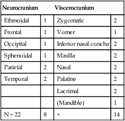
Skull
Neurocranium
Viscerocranium
Ethmoidal
1
Zygomatic
2
Frontal
1
Vomer
1
Occipital
1
Inferior nasal concha
2
Sphenoidal
1
Maxilla
2
Parietal
2
Nasal
2
Temporal
2
Palatine
2
Lacrimal
2
(Mandible)
1
N = 22
8
+
14

Major Sutures of the Skull.
Internal Features of Base of Skull.
Foramina of Skull.
Foramen/Opening
Bone
Structures Transmitted
Optic canal
Lesser wing of sphenoidal bone
Optic nerve
Ophthalmic artery
Sympathetic plexus
Superior orbital fissure
Greater and lesser wings of sphenoidal bone
Lacrimal nerve (V1)
Frontal nerve (V1)
Trochlear nerve (IV)
Oculomotor nerve (III)
Abducent nerve (VI)
Nasociliary nerve (V1)
Superior ophthalmic vein
Inferior orbital fissure
Between greater wing of sphenoidal bone and zygomatic
Infra-orbital vein
Infra-orbital artery
Infra-orbital nerve
Foramen spinosum
Greater wing of sphenoidal bone
Middle meningeal artery and vein
Foramen rotundum
Greater wing of sphenoidal bone
Maxillary division trigeminal nerve (V3)
Foramen ovale
Greater wing of sphenoidal bone
Mandibular division trigeminal nerve
Lesser petrosal nerve
Foramen lacerum
Between temporal bone (petrous area) and sphenoidal bone
Internal carotid artery
Foramen magnum
Occipital bone
Medulla oblongata
Vertebral artery
Meninges
Spinal roots of accessory nerve
Hypoglossal canal
Occipital bone
Hypoglossal nerve (XII)
Jugular foramen
Between temporal bone (petrous area) and occipital bone
Glossopharyngeal nerve (IX)
Vagus nerve (X)
Accessory nerve (XI)
Inferior petrosal sinus
Sigmoid sinus
Posterior meningeal artery
Mandible
Temporomandibular Joint
Anatomical Points
Clinical Points
Skull (Calvaria) Fractures
Le Fort Fractures
1.3 Superficial Face
Guide
Face
Scalp
Vascular Supply of the Face


