Patient Story
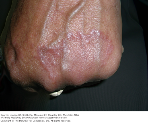
A 39-year-old woman presents with raised rings on her right hand only. Not knowing the correct diagnosis, another physician prescribed topical steroids and antifungal medicines with no benefit. The diagnosis of granuloma annulare (GA) was made by the typical clinical appearance and the patient was offered intralesional steroids. Triamcinolone acetonide was injected as seen in Figure 173-1, A. The patient noted improvement over the subsequent weeks, but within a month new lesions began to appear on her other hand (Figure 173-1, B). Additional injections were provided and 1 month later the patient had regression of the treated lesions but had new lesions on the right arm (Figure 173-2, A). At the next visit, the patient had new lesions on her feet as well (Figure 173-2, B). The diagnosis of disseminated GA was made and systemic treatment was started.
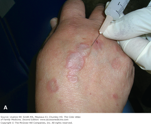
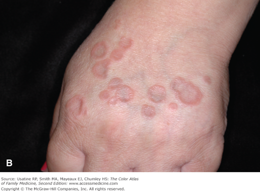
Figure 173-2
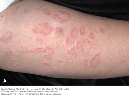
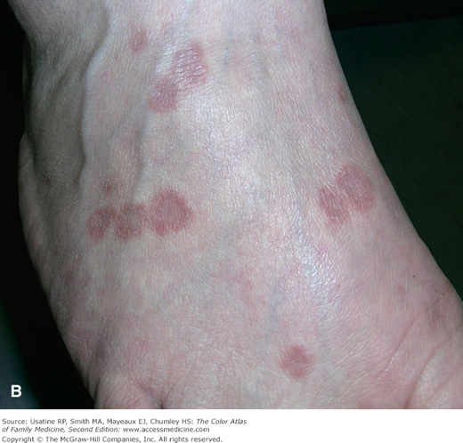
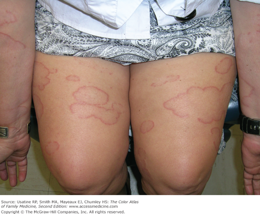
A. Same patient as in Figure 173-1 1 month later with new crops of lesions on the arms and feet. She has disseminated GA. Note the central area of hypopigmentation secondary to a previous steroid intralesional steroid injection. B. Disseminated GA on the foot of the same patient. The rings are flatter and many are conjoined. (Courtesy of Richard P. Usatine, MD.)
Introduction
Epidemiology
Etiology and Pathophysiology
- Benign cutaneous, inflammatory disorder of unknown origin.1
- Disease may be self-limiting, but may persist for many years.
- Reported associations include diabetes mellitus, viral infections (including HIV), Borrelia and streptococcal infections, insect bites, lymphoma, tuberculosis, and trauma.2,3
- One proposed mechanism for GA is a delayed-type hypersensitivity reaction as a result of T-helper-type cell (Th)-1 lymphocytic differentiation of macrophages. These macrophages become effector cells that express tumor necrosis factor (TNF)-α and matrix metalloproteinases. The activated macrophages are responsible for dermal collagen matrix degradation.4
- An association between high expression of gil-1 oncogene and granulomatous lesions of the skin, including GA, has been established.5
Risk Factors
Diagnosis
Annular lesions have raised borders that are skin-colored to erythematous (Figures 173-1 and 173-2). The rings may become hyperpigmented or violaceous (Figure 173-2, B). There is often a central depression within the ring. These lesions range from 2 mm to 5 cm. Although the classical appearance of GA is annular, the lesions may be arcuate instead of forming a complete ring (Figure 173-3). Most importantly, there should be no scaling as seen in tinea corporis (ringworm).

Each of the four types of GA has a different distribution. Localized and disseminated GA differ only in that disseminated lesions can spread to the trunk and neck and may be more pronounced in sun-exposed areas.6
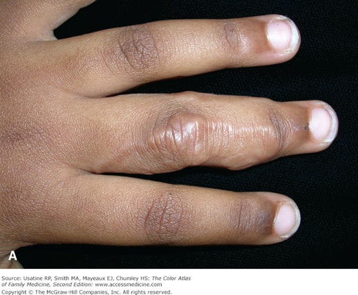
- Localized—This is the most common form of GA affecting 75% of GA patients.1 It typically presents as solitary lesions on the dorsal surfaces of extremities, especially of hands and feet (Figure 173-4).
- Disseminated or generalized—Adults are most affected by this form, which begins in the extremities and can spread to the trunk and neck (Figure 173-5).
- Perforating—Children and young adults present with 1 to hundreds of 1- to 4-mm annular papules that may coalesce to form a typical annular plaque. Although this form can appear anywhere on the body, it has an affinity for extremities, especially the hands and fingers.7 The papules may exude a thick and creamy or clear and viscous fluid.
- Subcutaneous—These lesions present as rapidly growing, nonpainful, subcutaneous or dermal nodules on the extremities, scalp, and forehead. Subcutaneous GA mainly affects children, with a mean age of 3.9 years (Figure 173-6).6 These lesions are often ill defined and less discrete.