Granular Cell Tumor

 Female predilection

image More prevalent in African Americans
• Wide anatomic distribution, usually arises in skin and subcutaneous tissue
image Head and neck region common (tongue, oral cavity)
• Usually solitary, painless nodule, plaque, or mass
image Multicentricity in 10% of cases
• Treatment: Complete surgical excision
• Excellent prognosis for benign lesions
• Malignant granular cell tumor (GrCT) (very rare) metastasizes in up to 50% of cases

Macroscopic

• Usually small (mean: 1-2 cm)

Microscopic

• Nonencapsulated, usually with irregular borders
• Overlying pseudoepitheliomatous hyperplasia in 30%
• Sheets, nests, and cords of plump, polygonal cells with abundant eosinophilic granular cytoplasm
• Typically prominent collagenous stroma
• Malignant GrCT shows ≥ 3 atypical features (pleomorphism, prominent nucleoli, increased mitoses, etc.)

Ancillary Tests

• PAS(+), diastase-resistant cytoplasmic granules
• Strong, diffuse S100(+), SOX10(+)
• Nuclear TFE3(+) in majority

Top Differential Diagnoses

• Congenital granular cell epulis
• Nonneural GrCT
• Hibernoma
• Melanoma
image
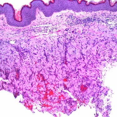
Granular Cell Tumor at Low MagnificationGranular cell tumor (GrCT) is a benign tumor of putative Schwann cell origin that occurs in a variety of anatomic sites but shows a predilection for the head and neck region, especially the tongue. This case presented as a dermal nodule on the chest wall, which was clinically suspicious for breast carcinoma.
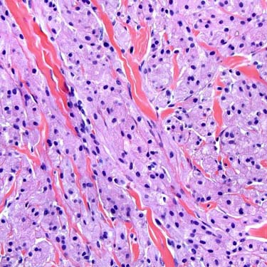
image
Sheets, Nests, and Cords of Tumor CellsThis tumor is composed of sheets, nests, and cords of plump, polygonal cells with abundant eosinophilic granular cytoplasm within a collagenous stroma.
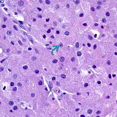
image
Prominent Granular CytoplasmCytologically, the cells of GrCT show prominent, granular cytoplasm, which represents numerous lysosomal structures. Nuclei are generally small and may be dark with dense chromatin or vesicular with a small nucleolus. Occasional scattered larger nuclei and nucleoli image may be seen in otherwise benign GrCT.
image
S100 ImmunohistochemistryStrong, diffuse S100 expression is characteristic of GrCT and can help to distinguish it from most entities in its differential diagnosis.

TERMINOLOGY

Abbreviations

• Granular cell tumor (GrCT)

Synonyms

• Granular cell myoblastoma
• Abrikossoff tumor
• Granular cell schwannoma

Definitions

• Benign tumor of putative schwannian origin composed of cells with abundant granular cytoplasm

CLINICAL ISSUES

Epidemiology

• Incidence
image Benign GrCT not uncommon
– Malignant GrCT extremely rare
• Age
image Any (peak: 40-60 years)
• Sex
image Female predilection
• Ethnicity
image More prevalent in African Americans

Site

• Wide distribution, usually arises in skin and subcutaneous tissue
image Rarely in deep soft tissue sites
• Head and neck region common
image Particularly tongue and oral cavity
• Also breast, bronchus, larynx, neurohypophysis, other viscera
• Rarely in nerves, lymph nodes

Presentation

• Usually solitary, slowly growing, painless nodule, plaque, or mass
image Multicentric in ~ 10% of cases
– May appear synchronously or metachronously
– May rarely arise in association with Noonan syndrome or LEOPARD syndrome
• Oral cavity/tongue lesions may show concurrent candidiasis

Treatment

• Complete surgical excision

Prognosis

• Overall excellent prognosis for benign lesions
image Local recurrence in < 10% of cases
• Multicentric tumors usually benign
• Malignant GrCT
image Up to 30% recur
image Up to 50% metastasize
image Adverse prognostic factors
– Local recurrence, metastasis
– Tumor size (> 5 cm), older patient age (> 50 years)

MACROSCOPIC

General Features

• Cutaneous lesions may show hyperplastic/verrucous appearance
• Cut surface firm, pale yellow, or cream colored

Size

• Usually small (mean: 1-2 cm)
image Deep lesions often larger (5-6 cm)

MICROSCOPIC

Histologic Features

• Nonencapsulated, usually with irregular borders
image May appear to infiltrate adjacent tissues (dermal collagen, adipose tissue, skeletal muscle)
– Tumor cells often appear to entrap small nerves
image Can extend directly up to surface epithelium
– May incite overlying pseudoepitheliomatous hyperplasia (30% of cases)
image Can mimic invasive squamous cell carcinoma in superficial biopsies
image Rare, plexiform growth pattern in dermis
• Sheets, nests, and cords of plump, polygonal cells
image 
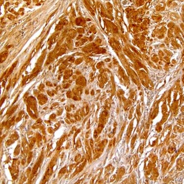
Abundant granular, eosinophilic cytoplasm
– Enlarged granules with halos (phagolysosomes) frequent (referred to as pustulo-ovoid bodies of Milian)
– Cell membrane often indistinct; appears syncytial
image Central small nuclei with variable dense to vesicular nuclei
– Small, subtle nucleoli may be present
– Mitoses absent
• Typically prominent collagenous stroma
image May be fibrotic or desmoplastic in longstanding lesions

Only gold members can continue reading. Log In or Register to continue

Stay updated, free articles. Join our Telegram channel

Apr 24, 2017 | Posted by in PATHOLOGY & LABORATORY MEDICINE | Comments Off on Granular Cell Tumor

Full access? Get Clinical Tree

Get Clinical Tree app for offline access