Glomus Tumors
Thomas Mentzel, MD
Key Facts
Terminology
Perivascular myogenic mesenchymal neoplasm composed of cells closely resembling smooth muscle cells of normal glomus body
Clinical Issues
Distal extremities, especially in subungual location
Typically small, red-blue, painful nodules
< 10% recur locally
Malignant glomus tumors highly aggressive
Macroscopic Features
Red-blue nodular lesions
Microscopic Pathology
Solid glomus tumor
Most common variant
Well-circumscribed nodular neoplasm
Small, uniform, round tumor cells
Centrally placed, sharply punched-out, round nuclei
Glomangioma
Comprises up to 20% of glomus tumors
Glomangiomyoma
Solid glomus tumor or glomangioma and elongated, spindled smooth muscle cells
Glomangiomatosis
Extremely rare variant
Malignant glomus tumor (glomangiosarcoma)
Exceedingly rare neoplasms
Enlarged size (> 2 cm) &/or subfascial/visceral location
Marked nuclear atypia
Increased number of mitoses
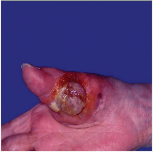 Clinical photograph shows a rare malignantglomus tumor involving the right thumb. |
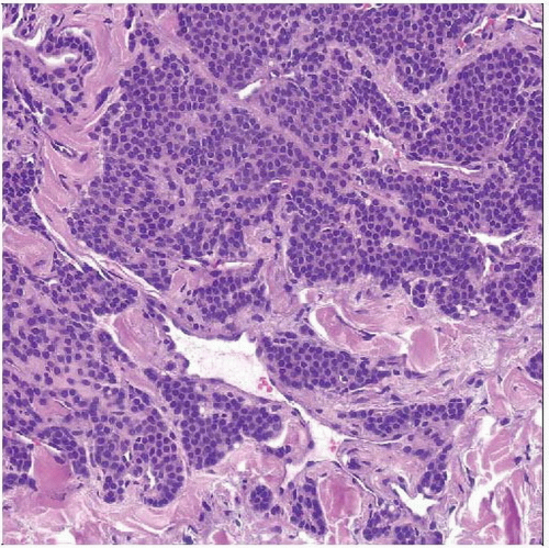 Hematoxylin & eosin shows perivascularly arranged myogenic tumor cells that contain uniform round nuclei in a benign glomus tumor. |
TERMINOLOGY
Abbreviations
Glomus tumor (GT)
Definitions
Perivascular myogenic mesenchymal neoplasm composed of cells closely resembling smooth muscle cells of normal glomus body
CLINICAL ISSUES
Epidemiology
Incidence
Rare
Account for < 2% of soft tissue neoplasms
Age
Predominantly in young adults
May occur at any age
Gender
No sex predilection
Site
Distal extremities
Often in subungual location
Rare in other anatomic locations (e.g., visceral organs, bone, mediastinum, nerve)
Skin, subcutis
Rare in deep soft tissue
Presentation
Painful mass
Typically small red-blue nodules
Long history of pain
Pain with exposure to cold &/or tactile stimulation
Usually solitary lesions
Rare multiple neoplasms
Multiple lesions more common in childhood
Natural History
< 10% recur locally
Malignant glomus tumors highly aggressive
Metastases and death of patients in up to 40% of cases
Treatment
Surgical approaches
Complete excision
Prognosis
Benign behavior in most cases
MACROSCOPIC FEATURES
General Features
Red-blue nodular lesions
MICROSCOPIC PATHOLOGY
Histologic Features
Perivascular myoid tumor cells
Small, uniform, round tumor cells
Centrally placed, sharply punched-out, round nuclei
Eosinophilic cytoplasm
Each cell surrounded by basal lamina
Predominant Pattern/Injury Type
Circumscribed
Predominant Cell/Compartment Type
Smooth muscle
Solid Glomus Tumor
Most common variant
Well-circumscribed nodular neoplasm
Contains numerous capillary-sized vessels
Nest of tumor cells surrounding capillaries
Stroma may show hyalinization
Stroma may show myxoid changes
Rare degenerative cytologic atypia
Rare vascular invasion
Peripheral rim of collagen (fibrous pseudocapsule)
May contain numerous hemangiopericytoma-like vessels
Rare oncocytic changes
Rare epithelioid variant
Glomangioma
Comprises up to 20% of glomus tumors
Most common type in patients with multiple lesions
Less well circumscribed
Stay updated, free articles. Join our Telegram channel

Full access? Get Clinical Tree


