Fundamental Concepts of Neoplasia: Benign Tumors and Cancer


Fundamental Concepts of Neoplasia: Benign Tumors and Cancer


The term neoplasia (from Greek, neo = new and plasis = a moulding) indicates the formation of new tissue or a tumor (from Latin for swelling) that may be benign or malignant. The primary task of diagnostic cytology is the microscopic diagnosis and differential diagnosis of malignant tumors or cancers and their precursor lesions. This chapter presents an overview of these groups of diseases that will attempt to correlate current developments in basic research with a description of morphologic changes observed in tissues and cells.


BRIEF HISTORICAL OVERVIEW

Cancer has been recognized by ancient Greeks and Romans as visible and palpable swellings or tumors, affecting various parts of the human body. In fact, the very name of cancer (from Greek, karkinos, and Latin, cancer = crab) reflects the invasive properties of the tumors that spread into the adjacent tissues and grossly mimic the configuration of a crab and its legs. Ancient Greeks were even aware that the prognosis of a karkinoma (carcinoma) of the breast was poor but also cited alleged examples of healing the disease by amputation. Over many centuries, numerous attempts were made based on clinical and autopsy observations to separate “tumors” caused by benign disorders, such as inflammation, from those that inexorably progressed and killed the patient, or true cancers. These distinctions could not be objectively substantiated until the introduction of the microscope as a tool of research. As was narrated in Chapter 1, the first recognition of microscopic differences between malignant and benign cells is attributed to Johannes Müller (1836). Müller’s work stimulated numerous investigators, including his student Rudolf Virchow, considered to be the founder of contemporary pathology, and led to the recognition of various forms of human cancer in the 19th century. The observations on microscopic makeup of cancer subsequently led to the recognition of precursor lesions
or precancerous states. The reader interested in the history of evolution of early human thoughts pertaining to cancer is referred to the books by M.B. Shimkin (1976), L.J. Rather (1978), and to the first chapter of this book.

In the first half of the 20th century, many attempts were made to shed light on the causes and sequence of events in cancer. Only a very few of these contributions can be mentioned here. As early as 1906, Boveri suggested that cancers are caused by chromosomal abnormalities. Differences in glucose metabolism between benign and cancerous cells were documented by Otto Warburg (1926), who believed that cancer was caused by insufficient oxygenation of cells or anoxia. Early measurements of cell components documented differences in nuclear and nucleolar sizes between benign and malignant lesions of the same organs (Haumeder, 1933; Schairer, 1935). The investigations of the sequence of events in experimental cancer supported the concept of two stages of development—initiation and promotion. The principal contributors of this theory were Friedwald and Rous (1944) and Berenblum and his associates (summary in 1974) who documented that cancer of the skin in animals (usually rabbits) may be produced more efficiently if the target organ, treated with a carcinogenic agent (such as tar) was treated with a second, noncarcinogenic agent, acting as a promoter. Knudson (1971, 1976) proposed the “two hit” theory of cancer, in reference to retinoblastoma, a tumor of the eye. The theory assumed that two events may be necessary for this cancer to occur—a genetic error that may be either congenital or acquired, followed by another carcinogenic event that again could be either genetic or acquired. With the discovery that the retinoblastoma gene (Rb gene; see below) is damaged or absent in some patients with retinoblastoma, the theory has proved to be correct. Subsequent developments in molecular studies of cancer led to the discovery of numerous tumor-promoting genes (oncogenes) and tumor suppressor genes, discussed later. It has been documented within recent years that the transformation of normal cells into cancerous cells is a multistep genetic process that is extremely complex. It is virtually certain today that carcinogenesis in various organs may follow different and, perhaps, multiple pathways. So far, there are only a very few genetic abnormalities that may represent common denominators of several cancers, such as the mutations of the p53 gene, discussed later, but the events preceding these mutations are in most cases still hypothetical and obscure.

None of these observations has shed much light on the morphologic and behavioral differences between cancer cells and benign cells, which are the principal topics of this book. Nonetheless, there is no further doubt that all tumors, whether benign or malignant, are genetic diseases of cells.


BENIGN TUMORS


Definition

Benign tumors are focal and limited proliferations of morphologically normal or nearly normal cells, except for their abnormal arrangement and quantity. Benign tumors may occur in any tissue or organ and are characterized by:


  • Limited growth


  • A connective tissue capsule


  • The inability to either invade adjacent tissue or metastasize


Classification

The most common benign tumors of epithelial origin are papillomas, usually derived from the squamous epithelium or its variants, such as the urothelium lining the lower urinary tract, and adenomas or polyps, derived from glandular epithelia (Fig. 7-1). Papillomas and polyps are visible to the eye of the examiner as pale or reddish protrusions from the surface of the epithelium of the affected organ. Microscopically, these tumors are characterized by a proliferation of epithelial cells, surrounding a core composed of connective tissue and capillary vessels. In some benign tumors of epithelial origin, such as fibroadenomas of the breast, the relationship of the epithelial structures and connective tissue is complex (see Chap. 29). Benign tumors may also originate from any type of supportive tissue (e.g., fat, muscle, bone) and usually carry the name of the tissue of origin, such as lipoma, myoma, or osteoma (Table 7-1).


Causes

The causes of benign tumors have not been fully elucidated but, in some of these tumors, chromosomal abnormalities have been observed (see Chap. 4 and Mitelman, 1991). The molecular significance of these abnormalities is not clear at this time. More importantly, Vogelstein and his group at Johns Hopkins observed that a tumor suppressor gene named APC (from adenomatous polyposis coli) is frequently mutated in benign polyps of patients with familial polyposis of colon, a disease characterized by innumerable colonic polyps and often leading to colon cancer. This gene
appears to interfere with adhesion molecules maintaining the normal integrity of colonic epithelium. The mutation of the APC gene may be a stepping stone to the development of colonic cancer (summary in Kinzer and Vogelstein, 1996). Although at this time no definitive information is available in reference to other benign tumors, it appears likely that they also occur as a consequence of mutations affecting genes essential in maintaining the normal relationship of cells.


Figure 7-1 Low-power view of rectal polyp. Note the central stalk of connective tissue and the benign glandular epithelium forming a mushroom around the stalk but also covering the stalk.


TABLE 7-1 CLASSIFICATION AND NOMENCLATURE OF HUMAN TUMORS*


Tissue Origin


Benign


Malignant (Cancer)


Stratified protective epithelium


Papilloma


Squamous or epidermoid carcinoma; urothelial carcinoma


Columnar epithelium, including that of glands


Adenoma or polyp


Adenocarcinoma, mucoepidermoid carcinoma
Occasionally epidermoid carcinoma


Mesothelia
Supportive tissues of mesodermal origin


Benign mesothelioma …omas according to the type of tissue involved (i.e., fatlipoma, bone-osteoma)


Malignant mesothelioma
Sarcoma (with designation of tissue type; i.e., liposarcoma, osteogenic sarcoma)


Lymphoid tissues


Hyperplasia


Malignant lymphomas


Blood cells


Leukemia


Tumors composed of several varieties of tissue


Benign teratomas


Malignant teratomas


*This simplified classification, although allowing a general orientation in tumor types, should not be taken too rigidly. A variety of malignant tumors may show a mixture of different types. Furthermore, combinations of sarcomas and carcinomas may occur. Special designations have been attached to a variety of benign and malignant neoplasms of some organs or systems. As the need arises, such diseases will be described in the text.


Another known cause of benign tumors is certain viruses. Thus, papillomaviruses may cause benign tumors in various species of animals. Certain types of the human papillomaviruses (HPVs) are the cause of benign skin and genital warts and papillomas of the larynx; other types, designated as “oncogenic,” are implicated in the genesis of cancer of the uterine cervix and other organs (see Chap. 11). It has been shown that some of the protein products, of the oncogenic types (which may also be involved in the formation of benign tumors), interact with protein products of genes controlling replication of DNA (p53) and the cell cycle (Rb) (see Chap. 11). No such information is available in reference to HPVs associated with benign tumors and the mechanisms of formation of warts remain an enigma at this time.


Cytologic Features

In general, the cells of benign epithelial tumors differ little from normal, although they may display evidence of proliferative activity in the form of mitotic figures. In general, the epithelial cells tend to adhere well to each other and form flat clusters of cells with clear cytoplasm and small nuclei, wherein cell borders are clearly recognized, resulting in the so-called honeycomb effect (Fig. 7-2).

Benign tumors of mesenchymal origin, such as tumors of fat (lipomas), smooth muscle (leiomyomas), or connective tissue (fibromas), can be sampled only by needle aspiration biopsies. In smears, the cell population resembles the normal cells of tissue of origin (i.e., fat cells, smooth muscle cells, or fibroblasts). As a warning, some malignant tumors of the same derivation may be composed of cells that differ little from their benign counterpart (see Chap. 24).

However, some benign tumors, such as tumors of endocrine
or nerve origin, may show significant abnormalities in the form of large, hyperchromatic, sometimes multiple nuclei that explain why the DNA pattern of such tumors may be abnormal (Agarwal et al, 1991). In the presence of such abnormal cells, the cytologic diagnosis of benign tumors may be very difficult. Benign tumors caused by human papillomaviruses, such as skin warts and condylomas of the genital tract or bladder, may show significant cell abnormalities that may mimic cancer to perfection.


Figure 7-2 Cells from a benign epithelial tumor. In this example from prostatic hyperplasia, there is a flat sheet of cells of nearly identical sizes. The cell borders among cells are recognizable as thin lines, giving the “honeycomb” effect.

Benign tumors of many organs show specific microscopic features that may allow their precise recognition, as will be discussed in detail in appropriate chapters. On the other hand, in some organs, such as the endometrium, the distinction between benign proliferative processes, known as atypical hyperplasia, and low-grade cancer may depend on the preference of the observer (see Chap. 14).


Behavior

Some benign tumors may regress spontaneously, such as skin warts. However, most benign tumors do not regress but achieve a certain size and then either stop growing or continue to grow at a very slow rate. Still, the size alone may interfere with normal organ function and may require removal. Other reasons for therapy may be necrosis or hemorrhage within the benign tumor that may cause acute discomfort to the patient. Also, a benign tumor may occasionally give rise to a malignant tumor although, on the whole, this is a rare event. The mechanisms and causes of such transformations are unknown, except for the colon, where it was shown, in high-risk populations, that a series of successive genetic abnormalities may lead from benign colonic polyps to cancer of the colon (see below).


MALIGNANT TUMORS (CANCERS)


Definition

Fully developed primary malignant tumors are characterized by several fundamental features that apply to all cancers:


  • Autonomous proliferation of morphologically abnormal cells results in abnormal, often characteristic tissue patterns and leads to the formation of a visible or palpable swelling or tumor.


  • Invasive growth involves growth of cancerous tissue beyond the boundaries of tissue of origin. The invasion may extend into adjacent tissues of the same organ and beyond.


  • Formation of metastases involves growth of colonies of cancer cells in distant organs, which again can proliferate in an autonomous fashion. For metastases to occur, the cancer cells must have the ability to enter either the lymphatic or blood vessels. Spread of cancer through lymphatics is known as lymphatic spread and leads to metastases to lymph nodes. Spread of cancer through blood vessels is known as hematogenous spread and may result in metastases to any organ in the body, whether adjacent to the tumor or distant (see Chap. 43).

The terms recurrent cancer and recurrence indicate a relapse of a treated tumor.


Classification

Cancers originating from epithelial structures or glands are known as carcinomas, whereas cancers derived from tissues of middle embryonal layer origin (such as connective tissue, muscle, bone) are classified as sarcomas. The names of yet other cancers of highly specialized organs or tissues may reflect their origin, for example, thymus = thymoma and mesothelium = mesothelioma. Cancers of blood cells are known as leukemias, and cancers of the lymphatic system as lymphomas (see Table 7-1).

Carcinomas and sarcomas may be further classified according to the type of tissue of origin, which is often reflected in the component cells. Carcinomas derived from squamous epithelium, or showing features of this epithelial type, are classified as squamous or epidermoid carcinomas. In this text, the term “squamous carcinoma” will be applied to tumors with conspicuous keratin formation, whereas tumors with limited or no obvious keratin formation will be referred to as “epidermoid carcinomas.” Carcinomas derived from gland-forming epithelium or forming glands are classified as adenocarcinomas. There are also carcinomas that may combine the features of these two types of cancer and are, therefore, known as adenosquamous or mucoepidermoid carcinomas. Carcinomas of highly specialized organs may reflect the tissue of origin, for example, hepatoma, a tumor of liver cells.

Sarcomas are also classified according to the tissue of origin, such as bone (osteosarcoma), muscle (myosarcoma), and connective tissue or fibroblasts (fibrosarcoma). Again, tumors derived from highly specialized tissues may carry the name of the tissue of origin, for example, glial cells of the central nervous system (glioma) or pigment-forming cells, melanoblasts (melanomas).

Yet other tumors may show combinations of several tissue types (hamartomas and teratomas), or reflect certain common properties, such as production of hormones (endocrine tumors). In certain age groups, tumors that show similar morphologic characteristics (although not cells of origin) have been grouped together as small-cell malignant tumors of childhood. The feature of all these tumors will be discussed in appropriate chapters.

Immunochemistry may be of significant help in classifying tumors of uncertain origin or type (see below and Chap. 45).


Risk Factors and Geographic Distribution

Only about 5% of all malignant tumors occur in children and young adults. Most cancers are observed in people past
the age of 50. In fact, it can be stated that advancing age is a risk factor for cancer. The reasons for this are speculative and most likely are based on reduced ability of the older organism to control genetic abnormalities that are likely to occur throughout the life of an individual but are better controlled in the younger age groups. A possible candidate is capping of chromosomes by telomeres that protect the ends of chromosomes from injury and that are reduced with age (de Lange, 2001). Another important risk factor is immunosuppression, particularly in patients with AIDS (Frisch et al, 2001).

Epidemiologic data from various continents and countries suggest that certain cancer types may preferentially occur in certain populations. For example, gastric cancer is very common in the Japanese, whereas cancer of the nasopharynx and esophagus is common in the Chinese. On the other hand, prostatic cancer is much less frequent in Japan than in the United States, where the disease is particularly common among African-Americans. Such examples could be multiplied. Epidemiologic studies have attempted to identify the causes of such events with modest success. It is known, for example, that among the Japanese living in Hawaii and the mainland United States, the rate of gastric carcinoma drops rapidly, and the change is attributed to a different diet. Several other environmental risk factors have been identified, but there are still huge gaps in our understanding of these events. The search for factors that may account for the geographic disparities is still in progress.


Causes

The first observations on the causes of human cancer had to do with environmental factors. Thus, an epidemic of lung cancer was observed in the 1880s in Bohemia (today the Czech Republic) in miners extracting tar that was subsequently shown to be radioactive (see Chap. 20). In the 1890s, after the onset of industrial production of organic chemicals, some chemicals were shown to cause bladder tumors (see Chap. 23). Asbestos has been linked with malignant tumors of the serous membranes (mesotheliomas; see Chap. 26), cigarette smoking with lung cancer, and exposure to ultraviolet radiation with skin cancers and melanomas. Many of these relationships have been studied by cancer epidemiology, a science that attempts to document in an objective, statistically valid fashion the relationship of various factors to cancer.

Another association of cancer is with infectious agents, such as viruses and bacteria (Parsonnet, 1999). Several RNA viruses, today known as retroviruses, have been shown to cause malignant tumors and leukemias in mice and other rodents, among them mammary carcinoma (Bittner, 1947; Porter and Thompson, 1948) and erythroleukemia in mice (de Harven, 1962). The ability of certain DNA viruses, such as the simian virus 40 (SV 40), to modify the features and the behavior of cells in culture has also been documented (Dulbecco, 1964). Such modified cells, when injected into the experimental animal, produce tumors capable of metastases.

In humans, a number of DNA viruses have been implicated in various malignant processes. As previously mentioned, human papillomaviruses (HPVs) of certain types have been linked with cancer of the uterine cervix (see Chap. 11) and the esophagus (see Chap. 24). Another DNA virus, the Epstein-Barr virus (EB virus) was implicated in Burkitt’s lymphoma and nasopharyngeal carcinoma. Virus of hepatitis B has been implicated in malignant tumors of the liver (hepatomas), whereas a newly discovered herpes virus type 8 has been found in association with vascular tumors, known as Kaposi’s sarcoma, and certain types of malignant lymphomas in patients with AIDS.

Bacteria, notably Helicobacter pylori, have now been implicated in the origin of gastric carcinoma and, perhaps, the uncommon gastrointestinal stromal tumors (GISTs) (see Chap. 24).

However, the vast majority of human cancers occur in the absence of any known risk factors. With the onset of molecular biology, the study of members of families with known high risk for certain cancers (cancer syndromes; see below) has led to the observations that they carried certain genetic abnormalities that were either recessive or dominant. These abnormalities have led to the studies of molecular underpinning of the events leading to cancer, discussed below.


Grading and Staging

Grading of cancers is a subjective method of analysis of cancers that attempts to describe the histologic (and sometimes cytologic) level of deviation from normal tissue or cells of origin. Grading is expressed in Roman numbers or equivalent phrases. If the histologic pattern of a cancer resembles closely the makeup of the normal tissue, and is composed of cells that closely resemble normal, it may be graded as well differentiated, or grade I. On the other extreme are cancers that barely resemble the tissue of origin, if at all, and are composed of cells that differ significantly from normal; such cancers can be classified as poorly differentiated, or grade III. Most cancers fall somewhere between the two extremes and are therefore classified as moderately well differentiated, or grade II. There are also systems of grading based exclusively on the configuration of nuclei of cancer cells, particularly in breast cancer (see Chap. 29). Several objective methods of measurements of cancer cells and their nuclei have been introduced to replace subjective grading (review in Koss, 1982). Grading may have some bearing on behavior of cancer, inasmuch as poorly differentiated tumors may be more aggressive than well differentiated. Grading is most valuable as a modifier of cancer staging.

Staging of cancers is based on an internationally accepted code to assess the spread of cancer at the time of diagnosis. The TNM system includes tumor size and extent of invasion (T), the involvement of the regional lymph nodes by metastases (N), and the presence or absence of distant metastases (M). The T group is usually subclassified and ranges from Tis (tumor in situ) or To, indicating a cancer confined to the tissue or organ of origin, to T1, T2,
T3 and T4, indicating tumor size and, in some instances, the depth of invasion.

Clinical staging is based on the results of inspection and palpation, now usually supplemented by radiologic techniques, such as magnetic resonance imaging (MRI) or ultrasound. Pathologic staging is based on examination of tissues surgically removed from the patient. The pathologic stage of a tumor may be higher than the clinical stage because, on microscopic examination, spread of cancer may be discovered in tissues that were clinically not suspect of harboring disease. The TNM system (sometimes combined with grading) is particularly useful in assessing the prognosis. To tumors have a much better outcome than T3 or T4 tumors. Tumors without metastases have a better prognosis than tumors with metastases. The TNM system is very useful in comparing the results of treatment of various malignant diseases in different institutions.


Behavior

In principle, all invasive cancers, if untreated, should lead to the death of the patient. However, even in untreated patients, the behavior of cancers may be extremely variable; some types of malignant tumors progress very slowly and take many years to spread beyond the site of origin, whereas other cancers progress and metastasize very rapidly, such as some cancers composed of small, primitive cells. In experimental systems, arrest and regression of malignant tumors was accomplished by a variety of manipulations (e.g., Silagy and Bruce, 1970) or by replacement of damaged genes and chromosomes. There is no doubt that occasionally, but very rarely, a spontaneous regression of human cancer can occur. Gene replacement therapy, however, has not been successful to date in human cancer.

Although statistical data are available today in reference to prognosis of most tumor types, experience shows that the rules do not always apply to individual patients. Except for the recognition of some cancer types with notoriously rapid progression, the classification of tumors by histologic (or cytologic) types may have limited bearing on behavior that is sometimes dependent on the organ of origin. For example, patients with squamous carcinomas of the cervix have a generally better prognosis and live longer than patients with cancers of identical type of the esophagus. As a group, adenocarcinomas of the breast are likely to be more aggressive and produce metastases sooner and more frequently than adenocarcinomas of the endometrium. In most common cancers, the behavior is better correlated with tumor stage than histologic type or grade, although grading may be a modifier of staging. The behavior of tumors of the same stage but different grades may vary. Tumors of higher grade often behave in a more aggressive fashion.


PRECURSOR LESIONS OF HUMAN CANCER

Although the concepts of precursor lesions of cancers were proposed in the early years of the 20th century (see Chap. 1), the existence and significance of these processes was firmly established during the last half of the 20th century. It is now known, with certainty, that tumors of epithelial tissue origin or carcinomas are preceded by abnormalities confined to the epithelium (Fig. 7-3). All these precursor lesions were initially classified as carcinoma in situ, and are now subdivided into several categories with names such as dysplasia or intraepithelial neoplasia. Some of these lesions may be graded by numbers (grade I, II, or III); by
adjectives, such as “mild,” “moderate,” or “severe”; or, within recent years, as “low-grade” or “high-grade” lesions. The grading has been used to indicate the makeup of these lesions—that is, the degree of morphologic abnormality—when compared with normal tissue of origin. Lesions resembling closely the epithelium of origin, albeit composed of abnormal cells, are classified as “low grade.” Lesions showing less or little resemblance to the epithelium of origin, usually composed of small abnormal cells, are classified as “high grade.” The grading has some bearing on the behavior of the precursor lesions, although in practice it has a rather limited value and reproducibility, as will be set forth in appropriate chapters.


Figure 7-3 Carcinoma in situ (severe dysplasia) of colon. A. Low power view of normal (right) and abnormal (left) colonic epithelium. B,C. The differences between the makeup of benign glands (B) lined by mucus-producing cells with small nuclei, and the malignant epithelium (C) composed of cells with no secretory function, very large nuclei, and evidence of mitotic activity are shown.

The general characteristics of precursor lesions of carcinomas are as follows:


  • The lesions are confined to the epithelium of origin.


  • They are composed of cells showing abnormalities that are similar but not necessarily identical to fully developed cancers.


  • Their discovery is usually the result of a systematic search, usually by cytologic techniques (e.g., in the uterine cervix, lung, oral cavity, urinary bladder, or esophagus) or incidental biopsies (e.g., colon). Although some precursor lesions may produce clinical abnormalities visible to the eye, such as redness, they do not form visible tumors. The discovery of precursor lesions is one of the main tasks of diagnostic cytology.


  • The behavior of precancerous lesions is unpredictable. Some of these lesions are capable of progression to invasive cancer but the likelihood of progression varies significantly from organ to organ. For example, in the urinary bladder at least 70% to 80% of untreated precursor lesions (flat carcinomas in situ and related lesions) will progress to invasive cancer, whereas, in the uterine cervix, the likelihood of progression does not exceed 20% (see Chaps. 11 and 23). The data for other organs are not secure because the system of discovery of precancerous lesions is not efficient. It must be noted that molecular genetic studies of precancerous lesions of the urinary bladder disclosed the presence of abnormalities that may also be observed in fully developed cancer. Similar observations were made in the sequence of events leading to cancer of the colon.


Progression of Intraepithelial Lesions to Invasive Cancer

Epidemiologic studies have shown that, as a general rule, precursor lesions occur in persons several years younger than persons with invasive cancer of the same type. Hence, it is assumed that several years are required for an intraepithelial lesion to progress to invasive cancer. For invasion to take place, the cells of the precursor lesion must break through the barrier separating the epithelium from the underlying connective tissue and, hence, must breach the basement membrane. One of two possible events must be assumed:


  • The cells composing the precancerous lesions acquire new characteristics that allow them to breach the basement membrane.


  • The basement membrane becomes altered and becomes a porous barrier to the cells.

Although the molecular mechanisms of such events are unknown at this time, there is evidence that some of the genes involved in carcinogenesis affect the adhesion molecules on cell membranes (see below). This relationship, when unraveled, may explain the mechanisms of invasion. Another, as yet unexplored, possibility is that the basement membrane is breached by ingrowing or outgrowing capillary vessels, thus paving the way for cancer cells to escape their confinement.


CURRENT TRENDS IN MOLECULAR BIOLOGY OF HUMAN CANCERS


Overview of the Problem

Cancer is a disease of cells that escape the control mechanisms of orderly cell growth and acquire the ability to proliferate, invade normal tissues, and metastasize. It is generally assumed that cancer is a clonal disorder derived from a single transformed cell (see below). The fundamental research issue was to determine whether cancer was the result of stimulation of cell growth, damage to the mechanisms regulating normal cell replication, or both. Marx (1986) referred to this dilemma as the Yin and Yang of cell growth control, referring to the old Chinese concept of contradictory forces in nature.

There were several significant problems with the study of molecular events in cancer. One of them was the heterogeneity of cancer cells—the observation that few, if any, cancer cells were identical. This phenomenon of cancer cell diversity was extensively studied by Fidler et al (1982, 1985), who documented that, in experimental tumors in mice, some cancer cells were capable of forming metastases and others were not. It has also been known for several years that the number and type of chromosomal abnormalities increased with the progression of cancer, reflecting the genomic instability in the cancer cells (recent review in Kiberts and Marx, 2002). Nowell (1976), who studied this phenomenon in leukemia, called it clonal evolution. In cytogenetic studies of fully developed solid cancers, the number of chromosomes in individual cancer cells is often variable and other aberrations of chromosomes may also occur (see Chap. 4). It is not an exaggeration to state that advanced human cancer represents a state of genetic chaos. The diversity of cancer cells, even within the same tumor, made it very difficult to assess whether observed molecular genetic abnormalities had universal significance or were merely an incidental single event (recent reviews in Tomlison et al, 2000, and Hahn and Weinberg, 2002).

The type of material that was available to the basic science investigators also posed similar problems. Fragments of cancerous tissues available for such purposes were usually derived from advanced tumors that were likely to show a great deal of heterogeneity and genetic disarray. In vitro
culture of human cancers is technically difficult, and the cell lines derived therefrom usually represented a single clone of cells that is not necessarily representative of the primary tumor.

Further complications arose when DNA or RNA were extracted from such tissue samples for molecular analysis. Besides tumor cells, such tissues always contained an admixture of benign cells from blood vessels, connective tissue stroma, inflammatory cells, and remnants of the normal organ of origin. The question as to what constituted tumorspecific findings, rather than findings attributable to normal cells, was often difficult to resolve. Many of these difficulties persist.

Some solutions to these dilemmas came from several unrelated sources. One of them was the discovery of growthpromoting DNA sequences, known as oncogenes, and their precursor molecules, the protooncogenes, in an experimental system of transformed rodent cells. The protooncogenes and oncogenes could be isolated and sequenced. The search could now begin for matching sequences in the DNA extracted from normal human tissues and cancer. The protooncogenes and oncogenes and their role in cancer are described below.

Another breakthrough occurred with the study of the patterns of occurrence of retinoblastoma, an uncommon malignant tumor of the retina in children. Knudson (1971) anticipated that a fundamental genetic abnormality accounted for the familial pattern of this disease. This abnormality was subsequently identified as a deficiency or absence of a gene located on chromosome 13, which was named the retinoblastoma (Rb) gene (see below for further discussion). Similar studies of families with “cancer syndromes” were also conducted. Such families, described by a number of investigators (Gardner, 1962; Li and Fraumeni, 1969; summaries in Lynch and Lynch, 1993; Fearon, 1997; Varley et al, 1997; Frank 2001) were characterized by a high frequency of occurrence of cancers in various organs. The most important cancer syndromes are listed in Table 7-2. By a variety of techniques known as linkage analysis, the genetic abnormalities could be identified and the genes localized—first to chromosomes, then to segments of chromosomes, and, finally, to the specific location on the affected chromosome. The isolation and sequencing of such genes were an essential step in studying their function and interaction with other genes.


TABLE 7-2 MOLECULAR GENETICS OF SOME CANCER SYNDROMES PERTINENT TO DIAGNOSTIC CYTOLOGY


Syndrome


Tumor Suppressor Gene


Chromosomal Location


Clinical Significance—Target Organs


Cytologic Targets


Familial polyposis coli


APC


5 q


colon cancer


metastatic cancer (liver, effusions, etc.)


Hereditary retinoblastoma


RB 1


13 q


eye: retinoblastoma


primary or metastatic


bone: osteosarcoma


Breast cancer (less commonly ovarian and tubal cancer)


BRCA 1


17 q


breast, ovary


primary or metastatic


BRCA 2


13 p


breast, pancreas


primary or metastatic


Li-Fraumeni


p53


17 p


diverse malignant tumors


primary or metastatic


Multiple endocrine neoplasia (MEN 1)


MEN 1


11 q


tumors of endocrine organs [thyroid, parathyroid, adrenal, pancreas (islands of Langerhans), pituitary]


primary or metastatic


Multiple endocrine neoplasia (MEN 2)


RET*


10 q


thyroid: medullary carcinoma,


primary or metastatic


adrenal: pheochromocytoma


Renal ca (part of von Hippel-Lindau syndrome)


VHL


3 p


kidney: carcinoma


primary or metastatic


Wilms’ tumor


WT 1


11 p


kidney: Wilms’ tumor


primary or metastatic


Peutz-Jeghers syndrome


STK 11+


19 p


associated with minimal deviation endocervical adenocarcinoma


endocervical adenocarcinoma


Hereditary melanoma


P16


9 q


skin: malignant melanoma


metastatic tumors


CDK4


12 q


* oncogene
q = long arm of chromosome

+ inactivation of protein kinases
p = short arm of chromosome


Of special value in this research were 
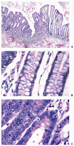
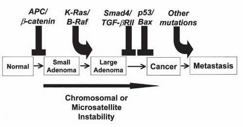
families with congenital polyposis of the colon, a disease process in which the patients develop innumerable benign colonic polyps and, unless treated, invasive cancer of the colon sooner or later. A group at The Johns Hopkins medical institutions in Baltimore, MD, led by Vogelstein, Fearon, and others, undertook a systematic study of genetic changes occurring
in benign colonic polyps, polyps with atypical features, early cancer, and invasive colonic cancer. These studies led to a model of carcinogenesis in the colon that postulated a sequence of genetic abnormalities leading from normal epithelium to polyps to cancer (Fig. 7-4). Although this model is not likely to be applicable to all cancers of the colon, let alone other organs, it stimulated a great deal of research on carcinogenesis. Perhaps the most important developments, resulting directly or indirectly from the studies of familial cancer, were the discovery of the role played by regulatory genes (tumor suppressor genes) in the events of cell cycle and the relationship of genes involved in cancer genesis with adhesion molecules that regulate the relationship of cells to each other and to the underlying stroma. These observations are discussed below.


Figure 7-4 Sequence of molecular events in the development of carcinoma of colon. (Courtesy of Dr. Bert Vogelstein, Johns Hopkins Medical Institutions, Baltimore, MD.)

Another development that proved to be of significance in this research was the Human Genome Project, which provided a great deal of information on the distribution of genes on human chromosomes. Although the map of the human genome has been completed and the significance and role played by most of the genes remain unknown, commercial probes to many of these genes have become available that allow the study of genetic abnormalities in various human cancers. The emerging information is, unfortunately, enormously complex and so far has shed little light on the initial events, or sequence of events, in solid human cancer. Still, the genome project led to the discovery of the human breast cancer genes BRCA1 and BRCA2, to be discussed below.


Figure 7-5 Schematic representation of the origin of an oncogene (sarcoma or src gene) in an experimental system in which malignant transformation of cultured cells is achieved by means of a retrovirus.


Protooncogenes and Oncogenes

The first significant observation shedding light on the molecular mechanisms of cancer was the discovery of oncogenes in the 1980s (summary in 
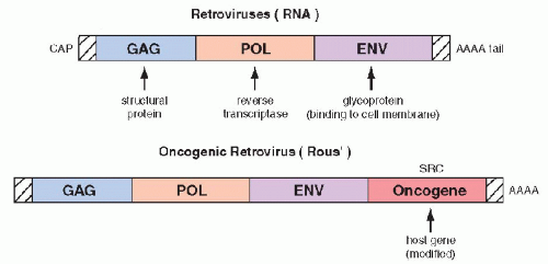
Bishop, 1987). The oncogenes were first identified in experimental systems in which cultured, benign rodent cells were infected with oncogenic RNA viruses (retroviruses) and were transformed into cells with malignant features. The viral RNA, by means of the enzyme reverse transcriptase is capable of producing cDNA that is incorporated into the native DNA (genome) of the cell, which becomes the source of viral replication. It has been observed that regulatory genes of host DNA, named protooncogenes, which may be incidentally appropriated by the viral genome, are essential in the transformation of the infected cells into cells with malignant features. The “stolen” host cell genes, when either overexpressed or modified (mutated), become a growth-promoting factor that has been named an oncogene (Fig. 7-5). The first oncogenes discovered were named ras (retrovirus-associated sarcoma or rat sarcoma). Several variants of the ras oncogenes were subsequently discovered and described with various prefixes, such as Kiras, Haras, and Nras, reflecting the initials of the investigators.

Shortly after the discovery of the first protooncogenes and oncogenes and their sequencing, their presence could be documented by Southern blotting and similar techniques in DNA from normal human tissues, in human tumors, and in cell lines derived therefrom. On the assumption that the study of oncogenes will provide the clue to the secrets
of abnormal cell proliferation in cancer, the search for other oncogenes and growth-promoting factors began in earnest and led to the discovery of a large number of such genes that have now been sequenced and traced to their chromosomal sites.

Two fundamental modes of oncogene function have been identified—overexpression (amplification) of a normal protooncogene product, and a 
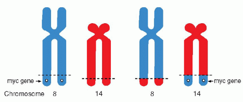
point mutation, a single nucleotide change in an exon of the gene, leading to a modified protein product. It is known that some oncogenes can be activated because their original chromosomal site has been disturbed by breakage and translocation of chromosomal segments, as observed in lymphomas and leukemias (see below). They may also be overexpressed in chromosomal fragments, such as C-myc oncogene, observed in the double-minute chromosomes of neuroblastoma (see Chap. 4).

The protooncogenes and the oncogenes exercise their activity through their protein products, many of which have been identified. For example, the genes of the ras family encode a group of proteins of 21,000 daltons, known as p21. Contrary to the initial hopes that all oncogenes would have a simple, well-defined function in the transformation of benign into malignant cells, it is now evident that the oncogenes are a diverse family of genes, with different locations within the cell and different functions. Several oncogenes have been traced to the nucleus (e.g., myc, myb, fos, jun), presumably interacting directly with DNA. Other proteins encoded by oncogenes have an affinity for cell membranes (e.g., ras, src, neu) or the cytoplasm (e.g., mos). These latter two groups of oncogenes appear to interact, on the one hand, with cytoplasmic and cell membrane receptors and, on the other hand, with enzymes, such as tyrosine kinase, that play a role in DNA replication. It is possible that the oncogenes located on cell membranes are instrumental in capturing circulating growth factors that stimulate proliferation of cells.

In solid human tumors, the activation or overexpression of various oncogenes has been shown to be a common event, unlikely to establish a simple cause-effect relationship between oncogene activation and the occurrence of human cancer. The presence of oncogene products could be demonstrated either by molecular biology techniques or by immunocytochemistry in many different human cancers. As an example, the presence of the ras oncogene product, p21, has been documented by us and others in gastric, colonic, and mammary cancer cells, and in several other human tumors (Czerniak et al, 1989, 1990, 
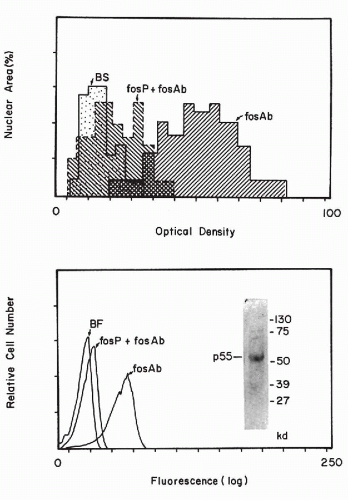
1992). In cytochemical studies, it was noted that oncogene products are variably expressed by cancer cells, some of which stain strongly and some that do not stain at all, suggesting heterogeneity of oncogene expression. It is possible that the expression of the oncogene products is, to some extent, cell cycle dependent (Czerniak et al, 1987). With image analysis and flow cytometric techniques (see Chaps. 46 and 47), the amount of the reaction product can be measured (Fig. 7-6). Press et al (1993) stressed that immunocytologic microscopic techniques with specific antibodies are probably more reliable in assessing the expression of an oncogene in tissues than is the Southern or northern blotting technique. The blotting techniques require the destruction of the tissue samples and, therefore, fail to provide information on the makeup of the destroyed tissue and on the proportion of normal cells in the sample.

However, there is no agreement on the diagnostic or prognostic value of such measurements in human solid tumors, with a few exceptions. For example, the elevated expression of the product of the oncogene HER2 (also known as c-erbB2), a transmembrane receptor protein, indicates poor prognosis and rapid progression of breast cancers in about 25% of affected women (Slamon et al, 1989). In fact, an antibody to the protein product of this gene has been developed commercially for human use and is of benefit in prolonging life in some women with advanced metastatic breast cancer (see Chap. 29). This is one of the first indications that knowledge of the oncogenes or tumorpromoting factors may be of benefit to patients. Although oncogenes play an important role in human cancer, their precise role is complex (summary in Krontiris, 1995). Weinstein (2002) suggested that individual cancers are “addicted” to their specific oncogenes and suggested that oncogene suppression may lead to cure.

As on example, the drug Gleevac (Novarrtis) has been shown to be effective against chronic myclogenous leukemia by blocking the oncogenic protein bcr = abl, the product of chromosome translocation.


Tumor Suppressor Genes and Gatekeeper Genes

The oncogene story became even more complicated with the identification of genes known collectively as tumor suppressor genes or gatekeeper genes. As previously mentioned, this research has been stimulated by studies of families with cancer syndromes (recent summary in Fearon, 1997; see Table 7-2). The first such gene discovered was the retinoblastoma (Rb) gene, located on the short arm of chromosome 13. Retinoblastoma is an uncommon, highly malignant eye tumor of childhood that occurs in two forms: (1) a familial form, in which usually both eyes are affected, and (2) a sporadic form, in which one eye is affected. Following treatment of retinoblastoma, other cancers, such as osteogenic sarcoma, may develop in the affected children. Thus, the defect of the Rb gene may have multiple manifestations.

It was postulated by Knudson in 1971 that retinoblastomas are the consequence of two mutational events (two-hit theory of cancer). The familial form of retinoblastoma implied a hereditary defect of some sort, supplemented by a single additional sporadic mutation, leading to cancer. In the sporadic form, two mutational events were anticipated against a normal genetic background. In retinoblastoma, the gene on chromosome 13 was frequently deficient or absent, thus fulfilling the first requirement of Knudson’s hypothesis. This gene has now been sequenced and its anti-tumor activity has been confirmed in vitro by Huang et al in 1988. It has been learned in recent years that the protein product of the Rb gene regulates the expression of one of the proteins regulating the cell cycle, known as D cyclins, which govern the transition of cells from G0 to G1 stage of
mitosis. It is postulated that the absence of, or damage to, the Rb gene deregulates the cell cycle, leading to cancer.


Figure 7-6 Measurement of fos p55 by computer-assisted image analysis (top) and flow cytometry (bottom). BS = background staining; fosP + fosAb = antibody to fos product p55 blocked by p55; fosAb = expression of unopposed antibody to p55; BF = background fluorescence. (Bottom right) Western blot of MCF7-KO protein extract incubated with antibody to c-fos p55. (Czerniak B, et al. Quantitation of oncogene products by computer-assisted image analysis and flow cytometry. J Histochem Cytochem 38:463, 1990.)

Another important regulatory gene is p53, a protein product of the gene located on the short arm of the chromosome 17 (Levine et al, 1991). p53 is a DNA binding protein that regulates the transcription of DNA, its repair by a cascade of other proteins, and is, therefore, considered to be a “guardian of the genome” (Lane, 1992). If a transcriptional error occurs, the replication is stopped until the error is repaired. The mechanism of arrest is mediated by a cell cycle inhibitor, protein p21WAF1/CIP1, which is different from the p21 protein of the ras gene. If the repair is not executed, the cell may enter into the cycle of programmed cell death or apoptosis, discussed in detail in Chapter 6.

The natural p53 product is short-lived and difficult to demonstrate; however, a gene mutation leads to a modified protein that has a much longer life span and can be demonstrated by a variety of techniques, including immunocytochemistry. Loss of heterozygosity of p53 (inactivation or mutation of one of the two identical genes within the cell) is a very common event in many human cancers of various organs, mainly in advanced stages (see later text). However, in some cancers, such as high-grade cancer of the endometrium, the mutation of p53 is presumed to occur as an early event (see Chap. 13). The presence of mutations of the Rb gene and of the p53 protein has been shown to confer a poor prognosis on some cancers, such as cancers of the bladder (Esrig et al, 1993; Sarkis et al, 1993), some malignant lymphomas (Ichikawa et al, 1997), and chondrosarcomas (Oshiro et al, 1998).

Other tumor suppressor genes include the recently identified breast cancer genes, BRCA1 and BRCA2 (see Table 7-2). The mutations of these genes have been observed in a larger proportion of Jewesses of Eastern European (Ashkenazi) origin than in other comparable groups of women (recent summary in Hofmann and Schlag, 2000). Although some of these women are at an increased risk for breast, and, to a lesser extent, ovarian and tubal cancer, and deserve close follow-up, the extent of risk for any individual patient cannot be assessed. In some of these women, preventive measures, such as a prophylactic mastectomy and oophorectomy have been proposed (Schraq et al, 1997). Clearly, many such dilemmas will occur as new risk factors for cancer are discovered. Silencing of tumor suppressor genes may be caused by methylation that does not involve DNA mutations (recent summary in Herman and Baylin, 2003).


Another set of genes involved in malignant transformation of normal cells into cancer cells is the susceptibility genes, considered by Kinzer and Vogelstein (1998) as “caretakers of the genome.” These genes, when mutated or inactivated, contribute indirectly to the neoplastic process, probably by regulating the relationship of the transformed cells to connective tissue stroma. Such genes have been observed in a colon cancer syndrome known as the hereditary nonpolyposis colorectal cancer (summary in Kinzer and Vogelstein, 1996). These observations bring into focus another critical issue in reference to cancer, namely the relationship of cancer cells to adhesion molecules that normally maintain order within the tissue and are critical in understanding the mechanism of cancer invasion and metastases. Several such molecules, such as cadherins (Takichi, 1991), integrins (Albelda, 1993), lamins (Liotta et al, 1984), and CD44 (Tarin, 1993), have been studied and have been shown to be of significance in cancer invasion and metastases.

It is the consensus of most investigators that cancer is a multistep process that includes sequential and progressive accumulation of oncogenes and inactivation of growth-regulating genes.


Microsatellite Instability

Another mechanism of cancer formation is instability of microsatellites, which are repetitive DNA sequences scattered throughout the genome. It has been noted that about 15% of colon cancers with a relatively normal chromosomal component display abnormalities of microsatellites (Gryfe et al, 2000; de la Chapelle, 2003). It is of note that the two pathways of colon cancer, i.e., chromosomal instability and microsatellite instability, result in different tumors with different behavior pattern and prognosis. The tumors with chromosomal instability are aneuploid, occur mainly in descending colon, and have a poor prognosis when compared with tumors with microsatellite instability, which tend to be diploid and occurring mainly in ascending colon (de la Chapelle, 2003).


Gene Rearrangement in Malignant Lymphomas and Leukemias: Effects of Translocations

Chromosomal abnormalities in leukemias have been studied since the onset of contemporary genetics. The Philadelphia chromosome (Ph), a shortened chromosome 22, described by Nowell and Hungerford in 1960 in chronic myelogenous leukemia, was the first documented chromosomal abnormality characteristic of any human cancer (see Chap. 4). With the availability of the techniques of chromosomal banding and molecular biology, the genetic changes in this group of diseases could be studied further. Many of these fundamental observations are of diagnostic and prognostic value. In many disease processes within this group of cancers, an exchange of chromosomal segments or translocation is observed (see Chap. 4 for a discussion of cytogenetic changes in human cancer). Thus, it has been shown that the Ph chromosome is the result of a translocation of portions of the long arm of chromosome 22 to the long arm of chromosome 9 [abbreviated as t(q9;q22)]. In certain forms of malignant lymphoma (notably in lymphomas of Burkitt’s type), there is a reciprocal translocation between segments of chromosomes 14 and 18 (Fig. 7-7).


Figure 7-7 Reciprocal translocation between fragments of chromosomes 8 and 14 in Burkitt’s lymphoma. The translation activates the myc gene and an adjacent immunoglobulin gene.

The results of a translocation can be:


  • Activation of a gene


  • Silencing of a gene


  • Formation of a novel protein by fusion of coding sequences of participating chromosomes

It is the last property that has served as a template for development of a new drug (Gleevec, Novartis) that is effective against the product of chromosomal translocation in chronic myelogenous leukemia. The new agent also appears to be active against a group of gastrointestinal tumors known as GIST (see Chap. 24).

Many genes affected by translocations have been localized, identified, and sequenced (Mitelman and Mertens, 1997). It is now known that the genes involved are often related to the principal sites encoding immunoglobulin genes. Adjacent genes often encode for certain oncogenes. For example, the 14:18 chromosomal translocation in B-cell lymphomas affects a gene known as bcl-2 and, in Burkitt’s lymphoma, the c-myc gene. Both the bcl-2 and c-myc genes have been shown to be inhibitors of programmed cell death or apoptosis and it is assumed that their mutation prevents apoptosis of genetically deficient cells and, thus, contributes to an unregulated proliferation of abnormal cells or cancer (Sanchez-Garcia, 1997).


Tumor Clonality: Loss of Heterozygosity

Another molecular feature that is common in cancer is loss of heterozygosity. The observation is based on the premise
that the two chromosomal homologues in each cell are not identical, as one is of paternal and the other of maternal origin. It is assumed that all cancer cells are derived from a single progenitor cell that carries the characteristics of only one parent and not both. One of the two genes may be inactivated or mutated. This phenomenon, known as loss of heterozygosity (LOH), could be first documented by studying the clonality of X chromosome expression in human cancer using markers to inactive chromosomal DNA. The most informative of these markers is X-linked human androgen receptor or Humara that can be effectively used in the detection of clonality of various disorders, whether malignant or benign (Willman et al, 1994). LOH can also be determined by Southern blotting searching for differences in expression of specific genes between the normal and malignant cells of the same person, using DNA amplified by polymerase chain reaction.


Angiogenesis

Another critically important factor in growth of cancer is supply of nutrients necessary to sustain the growth of cancer cells. A network of capillary vessels sustains the growth of cancer (Folkman and Klagsbrun, 1987). The molecules responsible for growth of capillaries have been identified and drugs directed against these factors are under development (Folkman, 1995). In the broad assessment of factors leading to cancer by Hahn and Weinberg (2002), angiogenesis is considered to be one of the five fundamental factors in the genesis of human cancer, the other four being resistance to growth inhibition, evasion of apoptosis, immortalization, and independence from mitotic stimulation.

In animal models, suppression of angiogenesis leads to regression of end-stage cancers (Bergers et al, 1999).


Immortality of Cancer Cells

In 1965, Hayflick pointed out that normal cells have a limited life span and die after 50 generations. These constraints are not applicable to cancer cells, which are theoretically immortal, as pointed out by Cairns (1975). Contrary to normal cells, given favorable conditions necessary for survival, cancer cells can live forever, and, in fact, they do so in tissue cultures. The reasons for the ability of cancer cells to proliferate without constraints are complex and not fully understood. One of the likely reasons is that cancer cells are deficient in control mechanisms protecting normal cells from faulty reproduction of DNA. In favor of this concept is the presence of the genetic defects, such as a mutated p53, in some cancer cells. This heritable defect in DNA control mechanisms may explain why the initial genetic changes lead to a cascade of events that result in ever increasing molecular (and chromosomal) disorders, discussed previously.

It is also possible that the chromosomes in cancer cells have a better mechanism of survival that prevents them from entering senescence, customary in normal cells. The guilty party may be the group of enzymes known as telomerases, enzymes governing the formation of telomeres, or the terminal endings of chromosomes (Blackburn, 1990). In normal cells, the length of the telomeres shrinks with age, presumably preventing the chromosomes from normal replication and leading to cell death after the 50 generations observed by Hayflick. Telomerases may be overexpressed in cancer and provide additional telomeres, thus preventing the senescence of chromosomes and leading to the immortality of cancer cells (Haber, 1995). Measuring the elevated expression of telomerase in cells has been used in the diagnosis of cancer (see Chap. 26).

The observations on the role of telomeres and telomerase in normal and cancerous cells are somewhat paradoxical; longevity of cells (and, by implications, multicellular organisms) and cancers have a common denominator. It is a matter for pure speculation at this time whether the efforts at extending the span of normal human life will inevitably lead to cancer. The same reasoning may, perhaps, be applied to the efforts at reversal of the malignant process by replacing damaged genes with intact genes. Such procedures have been repeatedly and successfully performed in vitro on tissue cultures but, so far, there is no reported evidence known to us of a successful application of such a procedure to multicellular organisms in vivo. It remains to be seen what long-term consequences this sort of a genetic manipulation of complex organisms may produce.


Animal Models

Many of the relationships among genes in cancer cells have been studied in experimental models in mice and rats wherein, by special manipulations on ova, certain genes can be removed or inserted. Knockout mice

Only gold members can continue reading. Log In or Register to continue

Stay updated, free articles. Join our Telegram channel

Jun 8, 2016 | Posted by in PATHOLOGY & LABORATORY MEDICINE | Comments Off on Fundamental Concepts of Neoplasia: Benign Tumors and Cancer

Full access? Get Clinical Tree

Get Clinical Tree app for offline access