Folic Acid1


Folic Acid1


Patrick J. Stover


HISTORICAL BACKGROUND

Also known as vitamin B9, vitamin Bc, vitamin M, Lactobacillus casei factor, folacin, and pteroyl-L-glutamic acid, folic acid was first discovered by Wills and Mehta in 1931 as a factor in yeast (“Wills’ factor”) that corrected the macrocytic anemia of pregnant Hindu women in India (1). The factor was later isolated from spinach leaves and was given the name folic acid (Latin folium, “leaf”) by Mitchell et al in 1941, who demonstrated that it was required for growth of Streptococcus lactis R (Streptococcus faecalis) (2). In 1945, the chemical synthesis of pure crystalline folic acid was reported in the journal Science (3). Synthetic folic acid was effective in reversing megaloblastic anemia that was refractory to treatment with liver extracts, but this agent was not able to prevent or improve neurologic damage that progressed from anemia that is now known to be caused by vitamin B12 deficiency.

Shortly after the discovery of folic acid as a growth-promoting factor, the development of folate antagonists as chemotherapeutics was undertaken by the Nobel Laureates Hitchings and Elion. In 1948, the folate antagonists aminopterin and, shortly thereafter, methotrexate were developed and administered to patients with childhood acute lymphoblastic leukemia and found to be effective treatments (4). This success led to the development of numerous anticancer and antimicrobial agents over the following 50 years that targeted folate-requiring enzymes. Beginning in the 1950s to the present time, folate-dependent enzymes were purified to homogeneity, biochemical pathways were elucidated, and later, their genes were cloned and structures determined. Beginning in the 1980s, the importance of folic acid in prevention of chronic diseases, certain cancers, and birth defects gained appreciation. This knowledge led to fortification of the food supply with folic acid in the United States, Canada, and other countries to prevent a class of common birth defects known as neural tube defects (NTDs). An excellent review of the history of folic acid was published in 2001 by Hoffbrand and Weir (1).


OVERVIEW OF FOLATE AND FOLIC ACID

Folate is a generic term that refers to a family of water-soluble B vitamins that are found in natural food and in biologic organisms (Fig. 26.1). Folates function as a family of enzyme cofactors that carry and chemically activate single carbons (referred to as one-carbons) for biosynthetic reactions. Folate is required for the biosynthesis of ribonucleotides and deoxyribonucleotide precursors for DNA synthesis. It is also required for amino acid metabolism, including the remethylation of homocysteine to methionine, and therefore functions in the regulation of gene expression by methylation. Hence, folate cofactors are
found in virtually all forms of life. Tetrahydrofolate (THF), which is the fully reduced form of the vitamin, carries one-carbons at one of three different oxidation levels ranging from methanol to formate (5, 6). The one-carbons are covalently bound to the N5 or N10 position of THF. In the cell, five different one-carbon substituted forms of THF are present: 10-formyl-THF; 5-formyl-THF; 5,10-methenyl-THF; 5,10-methylene-THF; and 5-methyl-THF, and each of these forms is interconverted in the cell through enzyme-mediated catalysis. Folates are also modified through the addition of a glutamate polypeptide that is polymerized through unusual γ-linked peptide bonds (7). The polyglutamate polypeptide increases the affinity of folate cofactors for folate-dependent enzymes and is required to retain folates within the cell and subcellular organelles. Folic acid (see Fig. 26.1) is not a biologically active form of folate, but it can serve as a provitamin because it is converted to the reduced, natural form of folate once transported into cells. It is an oxidized form of folate generated during the oxidative degradation of folate and normally does not accumulate in cells, although most degradation of THF is irreversible with degradation products that include oxidized pterin and para-aminobenzoyl-glutamate (8). Folic acid is also a synthetic form of folate present in fortified foods and in dietary supplements.


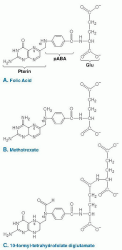
Fig. 26.1. The chemical structure of folic acid (A), methotrexate (B), and 10-formyl-tetrahydrofolate diglutamate (C). Folic acid contains a pterin ring that is bridged to para-aminobenzoic acid (PABA) through a methylene group to form pteroic acid. The addition of the glutamate residue (Glu) through a peptide linkage results in the formation of folic acid. Methotrexate (4-amino-10-methylpteroylglutamic acid) (B) is a folate analog, antagonist, and pharmaceutical agent that inhibits the activity of DHFR. Once transported into the cell, folic acid is reduced to tetrahydrofolate and is modified by the addition of a glutamate polypeptide containing up to nine glutamate residues linked by unusual γ-peptide linkages. THF is also modified by the addition of single carbons at the N5 or N10 position or that bridge the N5 and N10 positions. The carbon moieties are carried at the oxidation states of formate, formaldehyde, or methanol. The structure of 10-formyl-tetrahydrofolate diglutamate is shown in C.


DIETARY SOURCES

Folate is a vitamin and therefore must be acquired from the diet. Folate nutritional status is supported by the intake of the vitamin found in natural foods as well as dietary supplements and fortified foods (9). The best dietary sources of natural folate include fresh fruits, leafy green vegetables, yeast, liver, and legumes (10). Natural folates found in food are chemically labile and readily undergo irreversible oxidative degradation during food preparation and cooking. 5-Methyl-THF and formyl-substituted THF are the primary forms of folate present in natural foods, and they are also among the more stable forms of the vitamin. Folic acid, the synthetic, fully oxidized, and stable provitamin, is present in dietary supplements and fortified food (see Fig. 26.1). Folic acid has greater bioavailability than natural food folate because of its chemical stability and lack of a polyglutamate moiety, which impairs absorption across the intestinal epithelium (11). Once transported into the cell, folic acid is reduced to dihydrofolate (DHF) and subsequently THF by the enzyme DHF reductase (DHFR), and once fully reduced, folic acid is indistinguishable from natural food folate. Low levels of DHFR expression may result in the appearance of folic acid in the serum of individuals with high levels of folic acid intake (9). Evidence indicates that the total DHFR activity is highly variable among individuals—a finding that may indicate a variable capacity to metabolize folic acid among individuals (12).


RECOMMENDED DIETARY ALLOWANCES AND FOLIC ACID FORTIFICATION

The recommended intakes of folate established by the Food and Nutrition Board of the Institute of Medicine are shown in Table 26.1 (13). Dietary folate requirements are expressed as dietary folate equivalents (DFEs) because of the need to adjust for the increased bioavailability of folic acid compared with natural food folate (11). Folic acid is estimated to be 1.7 times more bioavailable than natural food folate. The recommended dietary allowance (RDA) for both men and women is 400 µg/day DFEs. The requirement for women of childbearing age is 400 µg of folic acid from fortified foods and supplements, in addition to food folate consumption from a varied diet (13). The tolerable upper intake level for adults was set at 1000 µg/day of folic acid exclusive of food folate, based on concerns that elevated intake of folic acid would exacerbate neurologic consequences of vitamin B12 deficiency.


TABLE 26.1 DIETARY REFERENCE INTAKES FOR FOLATEa


GROUP


INTAKE (µg OF DFE/d)


Infants (mo)


Adequate intake


0-5


65


6-11


80


Children and adolescents (y)


Recommended dietary allowance


1-3


150


4-8


200


9-13


300


14-18


400


Adults (y)


Recommended dietary allowance


≥19


400


Pregnant women


600


Lactating women


500


DFE, dietary folate equivalent.


aRequirements are expressed as dietary folate equivalents, to account for the increased bioavailability of folic acid compared with natural food folate.


Data from Food and Nutrition Board, Institute of Medicine. Folate. In: Dietary Reference Intakes for Thiamin, Riboflavin, Niacin, Vitamin B6, Folate, Vitamin B12, Pantothenic Acid, Biotin, and Choline. Washington, DC: National Academy Press, 1998:196-305; and Bailey LB, Gregory JF III. Folate metabolism and requirements. J Nutr 1999;129:779-82.


The United States and Canada mandated the addition of folic acid at a level of 140 µg/100 g of product to enriched flour in 1998 to achieve a predicted intake of 100 µg/day folic acid to reduce the incidence of NTDs (14). Before folic acid fortification, the median folate intake from food was estimated to be 250 µg/day. Population intake levels increased by 529 µg DFE/day between the interval 1998 to 1994 (before fortification) and 1999 to 2000 (after fortification) and then decreased by 135 mg between 1999 to 2000 and 2003 to 2004 (14). Folic acid fortification increased serum and red blood cell folate concentrations in the United States and decreased total plasma homocysteine levels by 6% to 13% (15, 16).


SITES OF INTESTINAL ABSORPTION

Folate absorption across the intestinal epithelium occurs in the acidic environment of the upper small intestine through the proton-coupled folate transporter (PCFT) (17), which was originally and probably incorrectly discovered as a heme transporter. Loss of PCFT function is associated with severe folate malabsorption, a finding indicating that it functions as the primary folate transporter in the gut. Only folate monoglutamates are bioavailable and absorbed. During digestion, the γ-glutamyl polypeptide of natural food folate is hydrolyzed to generate folate monoglutamate forms through a reaction catalyzed by the enzyme γ-glutamyl hydrolase. Folates circulate in serum as monoglutamate derivatives primarily in the form of 5-methyl-THF. In circulating red blood cells, 5-methyl-THF polyglutamates are the primary form of folates, although individuals with polymorphisms in the methylenetetrahydrofolate reductase (MTHFR) gene accumulate 10-formyl-THF in red blood cells (18). Transport into cells occurs primarily through the reduced folate carrier. Once transported into cells, folate monoglutamate derivatives are either converted to their polyglutamate forms by the addition of a γ-glutamyl polypeptide, usually consisting of five to nine glutamate residues in the cytoplasm or transported into mitochondria as monoglutamate derivatives and converted to polyglutamate forms in that compartment. The glutamate polypeptide serves to retain the vitamin within mitochondria and within the cell.


BIOLOGIC ROLES OF FOLATE

THF polyglutamates function as coenzymes that donate or accept one-carbons in an integrated network of biosynthetic and catabolic reactions involved in nucleotide and amino acid metabolism. Collectively, the network is commonly referred to as folate-mediated one-carbon metabolism. Folate metabolism is compartmentalized in the cytoplasm and nucleus (Fig. 26.2A) and mitochondria (Fig. 26.2B) (6). Each of these intracellular compartments is associated with specific metabolic pathways, and the compartments are interdependent through the exchange of common intermediates including formate, serine, and glycine (see Fig. 26.2B) (5, 19). Folate-mediated one-carbon metabolism also requires the water-soluble vitamins riboflavin (vitamin B2), niacin (vitamin B3), choline, pantothenic acid (vitamin B5), pyridoxal phosphate (vitamin B6), and cobalamin (vitamin B12) for its function (6) (see Fig. 26.2).


Cytoplasm

The de novo biosynthesis of purine and thymidylate nucleotides and the remethylation of homocysteine to methionine occur in the cytoplasm. The cytoplasm is the only compartment in the network that involves all the one-carbon substituted forms of THF (6). Formate serves as the primary source of one-carbon units for cytoplasmic one-carbon transfer reactions and is derived from amino acid catabolism in mitochondria (5, 20). In an adenosine triphosphate-dependent reaction, formate condenses with THF to form 10-formyl-THF, catalyzed by the 10-formyl-THF synthetase activity of the multifunctional enzyme methylenetetrahydrofolate dehydrogenase 1 (MTHFD1).


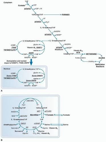
Fig. 26.2. Compartmentation of folate-mediated one-carbon metabolism in the cytoplasm, mitochondria, and nucleus. A. One-carbon metabolism in the cytoplasm is required for the de novo synthesis of purines and thymidylate and for the remethylation of homocysteine to methionine. One-carbon metabolism in the nucleus synthesizes thymidylate from uridylate and serine, and it occurs during the S phase of the cell cycle. B. One-carbon metabolism in the mitochondria is required to generate formate for one-carbon metabolism in the cytoplasm. The folate and amino acid carriers of the one-carbon unit are indicated by bold type. AdoHcy, S-adenosylhomocysteine; AdoMet, S-adenosylmethionine; ADP, adenosine diphosphate; ATP, adenosine triphosphate; DHF, dihydrofolate; DHFR, dihydrofolate reductase; DMGD, dimethylglycine dehydrogenase; dUMP, deoxyuridine monophosphate; GCS, glycine cleavage system; mFTHFS, mitochondrial formyltetrahydrofolate synthetase; mMTHFC, mitochondrial methenyltetrahydrofolate cyclohydrolase; mMTHFD, mitochondrial methylenetetrahydrofolate dehydrogenase; MTHFR, methylenetetrahydrofolate reductase; MTR, methionine synthase; M-tRNA-FT, methionyl-tRNA formyltransferase; NAD, nicotinamide adenine dinucleotide; NADP, nicotinamide adenine dinucleotide phosphate; NADPH, reduced nicotinamide adenine dinucleotide phosphate; Pi, inorganic phosphate; SD, sarcosine dehydrogenase; SHMT1, cytoplasmic serine hydroxymethyltransferase; THF, tetrahydrofolate; TYMS, thymidylate synthase.


Therefore, MTHFD1 is the primary entry point of one-carbons into the one-carbon metabolic network in the cytoplasm. The folate-dependent de novo synthesis of purine nucleotides involves 10 reactions and occurs through the formation of a multiple-enzyme complex termed a purineosome, which assembles when exogenous sources of purines are not available (21). The activated formyl moiety of 10-formyl-THF is incorporated into the number 2 and number 8 positions of the purine ring. In the third reaction of de novo purine biosynthesis, phosphoribosylglycinamide formyltransferase (GARFT) catalyzes the 10-formyl-THF-dependent conversion of glycinamide ribotide (GAR) to form formylglycinamide ribonucleotide (FGAR) and THF (see Fig. 26.2). In the ninth reaction, phosphoribosylaminoimidazolecarboxamide formyltransferase (AICARFT) catalyzes the 10-formyl-THF-dependent conversion of aminoimidazolecarboxamide ribotide (AICAR) to formylaminoimidazolecarboxamide ribonucleotide (FAICAR) and THF. Transformed cells are dependent on de novo purine biosynthesis, which accounts for the effectiveness of chemotherapeutic antifolates that target GARFT or AICARFT, including 6-R-dideazatetrahydrofolate (DDATHF; lometrexol that specifically targets GARFT) (22, 23, 24). Methotrexate (4-amino-10-methylpteroylglutamic acid) inhibits several folate-dependent enzymes, including both GARFT and AICARFT, by depleting 10-formyl-THF.

Alternatively, the one-carbon of 10-formyl-THF can be enzymatically reduced to 5,10-methylene-THF through the cyclohydrolase and reduced nicotinamide adenine dinucleotide phosphate (NADPH)-dependent dehydrogenase activities of MTHFD1. The de novo synthesis of thymidylate requires 5,10-methylene-THF as the one-carbon donating cofactor. 5,10-methylene-THF and uridylate are converted to thymidylate and DHF in a reaction catalyzed by the enzyme thymidylate synthase (TYMS). For this reaction, 5,10-methylene-THF serves both as a one-carbon donor and also as a source of two electrons through the oxidation of THF to DHF. THF is regenerated from DHF in a reaction catalyzed by the NADPH-dependent enzyme DHFR. To complete the de novo thymidylate synthesis cycle, THF is concerted to 5,10-methylene-THF by the three catalytic activities of MTHFD1, as described earlier, or alternatively by the vitamin B6-dependent enzyme serine hydroxymethyltransferase (SHMT1 and SHMT2α). The SHMT isozymes catalyze the conversion of serine to glycine to generate 5,10-methylene-THF from THF (see Fig. 26.2) (25). Several chemotherapeutic agents that target TYMS have been developed, including the fluoropyrimidines 5-fluorouracil (5-FU) and 5-fluoro-2-deoxyuridine (FdUrd), and the antifolates raltitrexed, pemetrexed, and methotrexate. These agents have been proven effective in the treatment of head, neck, breast, stomach, and colon cancers (26). These agents decrease TYMS catalytic function while also increasing cellular TYMS concentrations (27, 28) by preventing TYMS from binding to its mRNA or by decreasing the rate of ubiquitin-independent enzyme degradation (29, 30).

The remethylation of homocysteine to methionine occurs through folate-dependent and folate-independent pathways. For the folate-dependent pathway, 5,10-methylene-THF is reduced to 5-methyl-THF in a reaction catalyzed by the NADPH- and flavin adenine dinucleotide (FAD)-dependent enzyme MTHFR. 5-Methyl-THF is a cofactor for homocysteine remethylation to methionine, which is catalyzed by methionine synthase (MTR) in a vitamin B12-dependent reaction that converts 5-methyl-THF and homocysteine to methionine and THF. Homocysteine can be converted to methionine in a folate-independent reaction catalyzed by the enzyme betaine homocysteine methyltransferase, a reaction in which betaine serves as the one-carbon donor. Once formed, methionine can be adenosylated to form S-adenosylmethionine (AdoMet), which is a cofactor and one-carbon donor for numerous other methylation reactions (31). S-Adenosylhomocysteine (AdoHcy) is a product of AdoMet-dependent transmethylation reactions and is cleaved to form adenosine and homocysteine, which completes the homocysteine remethylation pathway.

These three metabolic pathways in the cytoplasm are highly interconnected and interdependent. Folate-dependent enzymes bind folate polyglutamate cofactors tightly with binding constants in the low micromolar or nanomolar range. The cellular concentration of folate-binding proteins exceeds that of folate derivatives (which are present at 25 to 35 µM), and therefore, the concentration of free folate in the cell is negligible (8, 32, 33). Consequently, independent of their origin, metabolic impairments of one-carbon metabolism rarely affect a single pathway, but rather they influence the entire network. This primarily occurs because the folate-dependent pathways compete for a limiting pool of folate cofactors in the cytoplasm (8, 34).

Only gold members can continue reading. Log In or Register to continue

Stay updated, free articles. Join our Telegram channel

Jul 27, 2016 | Posted by in PUBLIC HEALTH AND EPIDEMIOLOGY | Comments Off on Folic Acid1

Full access? Get Clinical Tree

Get Clinical Tree app for offline access