Chapter 33 First Trimester Ultrasound
Common indications
In the first trimester, the transvaginal transducer can be used to diagnose a living embryo, rule in intrauterine pregnancy, rule out multiple gestation, and calculate the gestational age (Figure 33-1).
Key steps
1. Position patient and prepare the transducer: The patient is asked to empty her bladder and place herself in the lithotomy position. Transvaginal ultrasound probes are useful to visualize the pregnancy in the first trimester. (See Chapter 34 on the second and third trimester ultrasound.) Apply a small amount of ultrasound gel to the inside of the probe cover and additional sterile lubricating gel to the tip of the covered transducer (Figure 33-2).
2. Introduce the transducer: Gently and slowly insert the transducer 3 to 4 cm in the sagittal plane. Keep the transducer orientation mark on the top (Figure 33-3). Gently move the transducer tip beyond the bladder, and place it near the uterus (Figure 33-4).
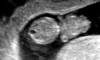
3. Identify the pelvic and fetal anatomy: Identify the cervix, the endocervical canal, and the decidua if present (Figure 33-5). Keeping the transducer in the sagittal plane, sweep it gently from left to right. Identify the black gestational sac surrounded by white decidua, and locate the fetus (Figure 33-6). Sweep through the entire uterus to rule out multiple gestations.
4. Fetal cardiac activity: Use the motion mode to extend a single line of ultrasound signal over time, to document fetal cardiac activity, and to determine the fetal heart rate (Figure 33-7). Failure to identify an intrauterine embryo, gestational sac, or yolk sac can indicate an ectopic gestation. If the quantitative βhCG is appropriately elevated, an empty uterus is an ectopic pregnancy until proven otherwise.
Stay updated, free articles. Join our Telegram channel

Full access? Get Clinical Tree


