Patient Story
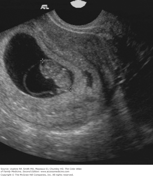
A 22-year-old woman presents with no menstrual period for approximately 2 months (she has irregular menses.) She is complaining of morning sickness but is otherwise feeling well. A urine pregnancy test confirms she is pregnant. Figure 77-1 shows a fetus of 9 weeks estimated gestational age (EGA).
Introduction
Obstetrical ultrasound has become a vital tool in our ability to properly care for the pregnant patient. Vast technologic improvements have made visualization of the pregnancy even better and improved our diagnostic capabilities, ranging from the normal pregnancy to the extremely early ectopic pregnancy. Ultrasonography (US) allows for a relatively detailed assessment of fetal gestational age, development, number of fetuses, and anatomy in utero. Most pregnancies in the United States undergo ultrasound imaging for various indications.
Epidemiology
- Women who receive antenatal care have lower maternal and perinatal mortality and better pregnancy outcomes.1 However, the optimal components of prenatal care have not been rigorously examined in well-designed studies.
- In the United States in 2003, 84.1% of pregnant women obtained prenatal care in the first trimester, and only 3.5% received no care or initiated prenatal care in the third trimester.
Etiology and Pathophysiology
- Ultrasound is used to estimate gestational age and to calculate the expected date of delivery (EDD). Ultrasound is especially helpful when menses are irregular, the last menstrual period (LMP) is unknown, or in patients who conceived while using hormonal contraceptives. A Cochrane review noted with more accurate dating there was a reduction in intervention for postterm pregnancy.2
- The Routine Antenatal Diagnostic Imaging with Ultrasound (RADIUS) trial was a randomized trial of routine obstetrical ultrasound screening.3

Stay updated, free articles. Join our Telegram channel

Full access? Get Clinical Tree


