and Natalia Buza1
(1)
Department of Pathology, Yale University School of Medicine, New Haven, CT, USA
Keywords
Serous tubal intraepithelial carcinoma (stic)High-grade serous carcinomaMetastatic tumors of fallopian tubeTubal epithelial hyperplasiaEmbryonic remnants/cystsEndometriosisSalpingitisEctopic pregnancyRisk-reducing salpingo-oophorectomyIntroduction
Neoplastic or reactive lesions presenting at the fallopian tube as a primary site of involvement are uncommon. The fallopian tube is most often received for intraoperative frozen section evaluation as part of a salpingo-oophorectomy specimen. The primary disease site may not always be accurately determined by preoperative imaging studies or the surgeon’s intraoperative assessment, and the specimen sent to pathology as an ovarian/adnexal mass occasionally may reveal a primary fallopian tube disease process. The most common primary fallopian tube lesions include epithelial neoplasms—benign, borderline, or malignant—embryonal remnants and cysts (e.g., paratubal cyst), endometriosis, inflammatory conditions, and ectopic pregnancy. Frozen section of the fallopian tube is indicated when the lesion is located within the tube or the primary site—ovarian versus tubal—cannot be determined with certainty based on the gross examination. Thorough intraoperative gross evaluation of the fallopian tube is typically sufficient when the main lesion is of primary ovarian or uterine origin. The role and optimal pathology protocol for intraoperative assessment of risk-reducing salpingo-oophorectomy specimens is still evolving, and currently no standardized guidelines exist. This chapter covers the frozen section pathologic features and implications of the most common neoplastic and nonneoplastic tubal lesions and clinicopathologic considerations of intraoperative evaluation of risk-reducing salpingo-oophorectomy specimens.
Primary Tumors of the Fallopian Tube
Serous Adenofibroma
Most often an incidental finding.
Small, benign biphasic tumor, typically located at the fimbriated end of the fallopian tube [1].
May form a grossly recognizable mass.
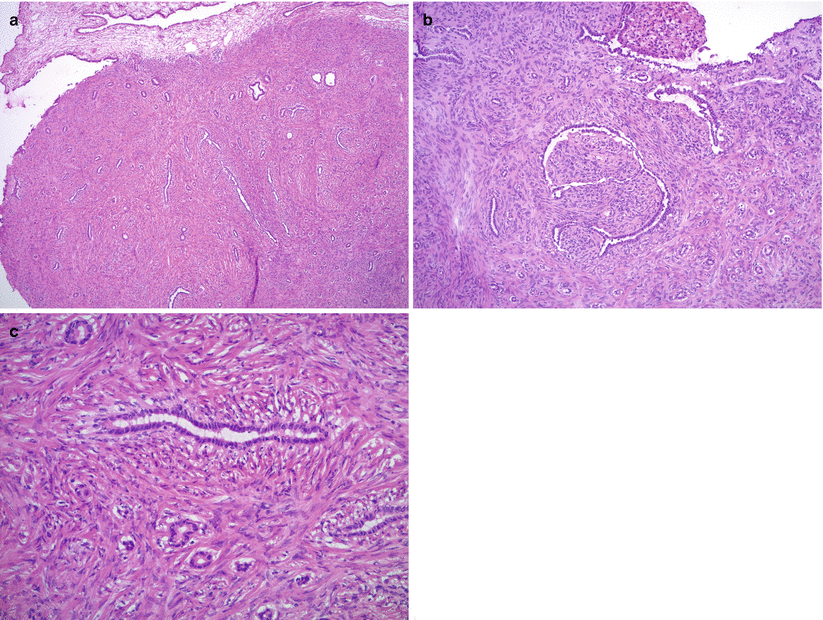
Histology is similar to its ovarian counterpart: variably sized irregular glands lined by benign tubal-type epithelium and fibromatous stromal component (Fig. 7.1).

Fig. 7.1
Serous adenofibroma. Variably sized, round, or irregularly shaped glands in a background of fibromatous stroma; note the adjacent tubal fimbria (a, upper left portion of image). The glands are lined by a single layer of tubal-type epithelium without atypia (b, c)
No significant nuclear atypia or mitotic activity.
Differential diagnosis
Serous borderline/atypical proliferative tumor (SBT/APST)
Diagnostic pitfalls/key intraoperative consultation issues
Lack of significant cytological atypia and papillary epithelial proliferation separates this entity from SBT/APST.
Serous Borderline/Atypical Proliferative Tumor (SBT/APST)
Rare tumor, histologically resembling its ovarian counterpart.
Clinicopathologic implications and prognosis are similar to ovarian SBT/APST.
Serous Tubal Intraepithelial Carcinoma (STIC)
Noninvasive serous carcinoma, most often arising in the fimbria or distal end of the fallopian tube.
Most STICs do not form a grossly recognizable mass lesion; however, rare cases of exophytic noninvasive serous carcinoma may be identified on gross examination as a small nodule [6].
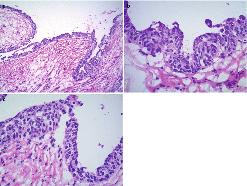
Microscopically STIC demonstrates multilayered epithelium with hyperchromatic, markedly atypical cells (Fig. 7.2).

Fig. 7.2
Serous tubal intraepithelial carcinoma (STIC). Multilayered epithelium (a) with loss of polarity, hyperchromasia, increased nuclear to cytoplasmic ratio, and severe cytological atypia are characteristic (b, c). Mitotic figures are common (c)
High nuclear to cytoplasmic ratio.
Prominent nucleoli.
Loss of polarity.
Absence of cilia.
Mitotic figures are often present.
Tumor cells may exfoliate into the tubal lumen.
Absence of stromal invasion.
Differential diagnosis
Tangential sectioning of tubal epithelium
Benign metaplastic changes—transitional, squamous, or papillary syncytial metaplasia—of the tubal epithelium
Tubal epithelial hyperplasia
Atypical epithelial lesions falling short of STIC morphologic criteria
Invasive tubal high-grade serous carcinoma
Secondary tubal spread from other gynecologic—ovarian or uterine—primaries
Diagnostic pitfalls/key intraoperative consultation issues
Atypical epithelial lesions falling short of STIC morphologic criteria do not need to be specified on frozen section as they do not require further surgery.
Peritoneal washing is recommended.
No uniform guidelines on the role and extent of surgical staging; approximately 50 % of patients with STIC in the literature underwent hysterectomy and omental biopsy/omentectomy, and only 24 % had lymph node sampling performed [7].
The diagnosis of STIC on frozen section (i.e., on morphology alone, without ancillary studies) may be very challenging, and a conservative approach or deferral to permanent sections is reasonable in difficult cases to avoid overdiagnosis.
Invasive tubal serous carcinoma typically requires surgical staging [8].
Secondary tubal involvement—by direct spread or metastasis—by other gynecologic primary tumors (ovarian or endometrial) may colonize the tubal epithelium mimicking STIC.
If the diagnosis of ovarian or endometrial malignancy is established (previously or intraoperatively), additional tubal involvement (whether secondary or synchronous primary tumor) typically does not have a significant impact on the surgical management.
Metastatic tumors from non-gynecologic primaries may also mimic STIC, but they usually involve the stroma of fallopian tube and multiple other organs as well.
In patients with prior history of non-gynecologic malignancy, morphologic comparison with the prior specimen(s)—if available at the time of frozen section—should be made.
High-Grade Serous Carcinoma (Invasive)
Most common type of tubal carcinoma.
May present with abdominal/pelvic pain or may be an incidental finding.
May involve bilateral fallopian tubes.
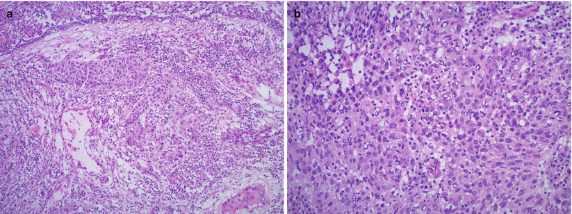
Histologically resembles high-grade ovarian serous carcinoma (see Chap. 8) (Fig. 7.3).

Fig. 7.3
High-grade serous carcinoma of the fallopian tube. Markedly atypical epithelial cells infiltrating the fallopian tube stroma in the form of solid nests and glandular structures with slit-like spaces. Note the overlying tubal epithelium (a). High nuclear to cytoplasmic ratio, prominent nucleoli, and frequent mitoses are seen (b)
Papillary, solid, or slit-like glandular pattern.
Marked nuclear atypia and frequent mitotic figures, including atypical mitoses.
Stromal invasion.
Necrosis and lymphovascular invasion may be apparent on frozen section.
Psammoma bodies may be seen.
Differential diagnosis
Serous tubal intraepithelial carcinoma (STIC)
Secondary tubal spread from other gynecologic—ovarian or uterine—or non-gynecologic primaries
Diagnostic pitfalls/key intraoperative consultation issues
Secondary tubal involvement—via direct spread or metastasis—by ovarian or endometrial serous carcinoma is common.
If the diagnosis of ovarian or endometrial malignancy is established (previously or intraoperatively), additional tubal involvement (whether secondary or synchronous primary tumor) typically does not have a significant impact on the surgical management.
Metastatic tumors from non-gynecologic primaries (most commonly breast or gastrointestinal) may mimic primary tubal carcinoma.
In patients with prior history of non-gynecologic malignancy, morphologic comparison with the prior specimen(s)—if available at the time of frozen section—should be made.
Adenomatoid Tumor
Benign tumor of mesothelial origin.
Usually small, incidental finding (<2 cm in size).
May be identified grossly as a solid, white, or yellow mass lesion.
Microscopic features are similar to its uterine counterpart (see Chap. 5).
Proliferation of small, irregular glandular spaces lined by bland mesothelial cells.
No significant nuclear atypia or mitotic activity.
Basophilic mucoid material may be seen in gland lumens.
Differential diagnosis
Angioma and lymphangioma
Stay updated, free articles. Join our Telegram channel

Full access? Get Clinical Tree


