Patient Story
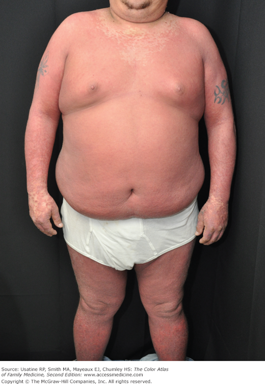
A 34-year-old man presented with red skin from his neck to his feet for the last month (Figure 156-1). He was having a lot of itching and his skin was shedding so that wherever he would sit, there would be a pile of skin that would remain. He denied fever and chills. He admitted to smoking and drinking heavily. The patient’s vital signs were stable with normal blood pressure and he preferred not to be hospitalized. He had some nail pitting but no personal or family history of psoriasis. The presumed diagnosis was erythrodermic psoriasis but a punch biopsy was done to confirm this. A complete blood count (CBC) and chemistry panel were ordered in anticipation of the patient needing systemic medications. A purified protein derivative (PPD) was also placed. The patient was started on total body 0.1% triamcinolone under wet wrap overnight and given a follow-up appointment for the next day. The patient was also counseled to quit smoking and drinking. The following day his labs showed mild elevation in his liver function tests (LFTs) only. The following day his PPD was negative and he was already feeling a bit better from the topical triamcinolone. Cyclosporine was started and the patient improved rapidly.
Introduction
Erythroderma is an uncommon condition that affects all age groups. It is characterized by a generalized erythematous rash with associated scaling. It is generally a manifestation of another underlying dermatosis or systemic disorder. It is associated with a range of morbidity, and can have life-threatening metabolic and cardiovascular complications. Therapy is usually focused on treating the underlying disease, as well as addressing the systemic complications.
Epidemiology
Erythroderma or exfoliative dermatitis is an uncommon condition that is generally a manifestation of underlying systemic or cutaneous disorders.
- It affects all age groups, from infants to the elderly.
- In adults, the average age of onset is 41 to 61 years of age, with a male-to-female ratio of 2:1 to 4:1.1
- It accounts for approximately 1% of all dermatologic hospital admissions.2
- It can be a very serious condition resulting in metabolic, infectious, cardiorespiratory, and thermoregulatory complications.3
Etiology and Pathophysiology
In almost 50% of cases, erythroderma occurs in the setting of a preexisting dermatosis; however, it may also occur secondary to underlying systemic disease, malignancy, and drug reactions. It is classified as idiopathic in 9% to 47% of cases.3
- The pathophysiology is not fully understood, but it is related to the pathophysiology of the underlying disease. However, the factors that promote the development of erythroderma are not well defined.
- The rapid maturation and migration of cells through the epidermal layer results in excessive scaling. The rapid turnover of the epidermis also results in fluid, electrolyte, and protein losses that may have severe metabolic consequences, including heart failure and acute respiratory distress syndrome.4
- The underlying pathogenesis may be an interaction of immunologic modulators, including Interleukins 1, 2, and 8, as well as tumor necrosis factor.2
Table 156-1 presents the conditions most commonly associated with erythroderma. Dermatologic conditions commonly associated with erythroderma include:1–5
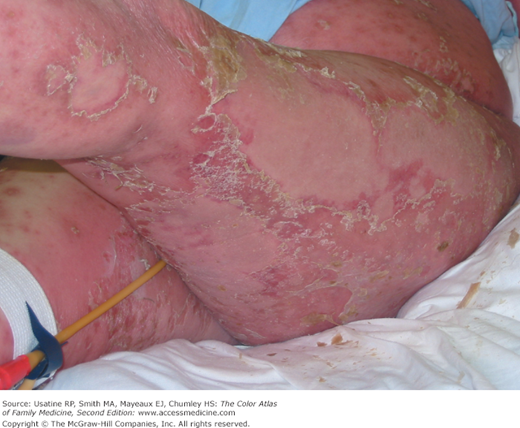
- Psoriasis, especially generalized pustular psoriasis with exfoliation (Figures 156-1, 156-2, and 156-3).
- Atopic dermatitis (Figure 156-4).2,3
- Contact dermatitis (Figure 156-5).
- Seborrhea (Figure 156-5).
- Pityriasis rubra pilaris.
- Bullous pemphigoid.4
- Impetigo herpetiformis.4
- Photosensitivity reaction.4
Dermatoses | Infections | Systemic/Cancer | Pediatric | Drugs |
|---|---|---|---|---|
Psoriasis (Figures 156-1, 156-2, 156-3, and 156-8) | HIV | Sarcoidosis | Omenn syndrome | Penicillins |
Atopic dermatitis (Figure 156-4) | Norwegian scabies | Thyrotoxicosis | Kwashiorkor | Sulfonamides |
Contact dermatitis (Figure 156-5) | Hepatitis | Graft versus host reaction | Cystic fibrosis | Tetracycline derivatives |
Seborrhea (Figure 156-5) | Murine typhus (Figure 156-6) | Dermatomyositis | Amino acid disorders | Sulfonylureas |
Pityriasis rubra pilaris | Human herpesvirus 6 | Lung cancer | Immunodeficiency | Calcium channel blockers |
Photosensitivity reaction | Toxic shock syndrome | Colon cancer | Captopril | |
Bullous pemphigoid | Staphylococcal scalded skin syndrome | Prostate cancer | Thiazides | |
Impetigo herpetiformis | Histoplasmosis Tuberculosis | Breast cancer B- and T-cell lymphoma (Figure 156-7) Leukemia | NSAIDs Barbiturates
Vancomycin Lithium Antivirals Antimalarials |


