Patient Story
A 57-year-old farm worker presents with itchy red rings on his body that have come and gone for more than 13 years (Figures 206-1 and 206-2). The erythematous annular eruption was visible on his abdomen, legs, and arms. Figure 206-2 shows the typical “trailing scale” of erythema annular centrifugum (EAC). A KOH preparation was negative for fungal elements and the patient was given the diagnosis of EAC. He recently began using paint thinner to “dry out the rash” and decrease the itching. Because topical steroids did not provide any relief for him in the past, we offered the option of using calcipotriol ointment. He chose to try the calcipotriol and stop using paint thinner.
Figure 206-1
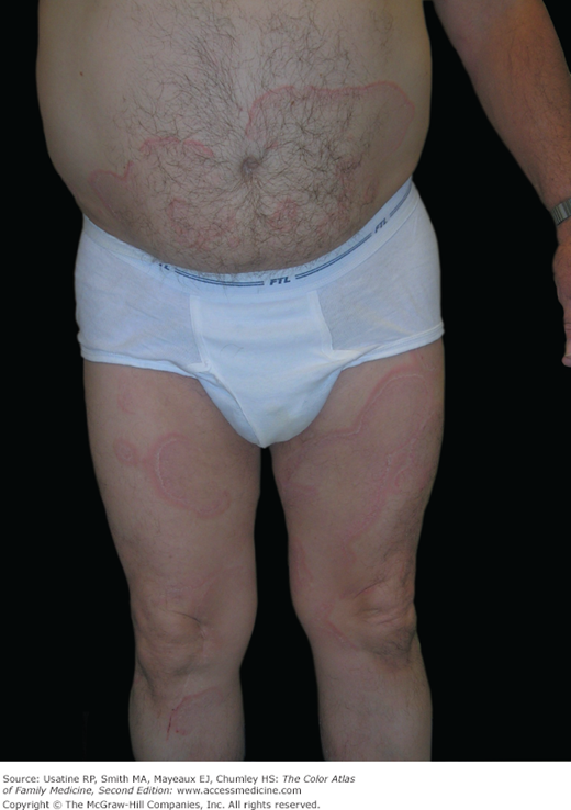
Erythema annulare centrifugum with large erythematous rings on the trunk and legs of a 57-year-old man. (Courtesy of Richard P. Usatine, MD. Reproduced with permission from Brand ME, Usatine RP. Persistent itchy pink rings. J Fam Pract. 2005;54(2):131-133. Reproduced with permission from Frontline Medical Communications.)
Figure 206-2
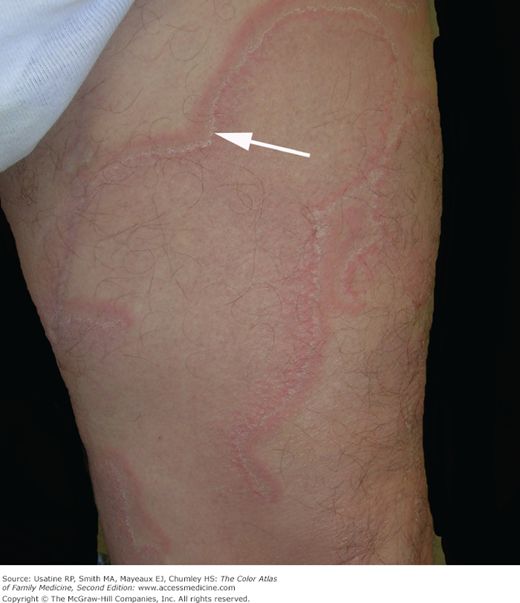
Erythema annulare centrifugum with conjoined rings on the thigh. Arrow pointing to “trailing scale,” which appears as a white scaling line within the erythematous border. (Courtesy of Richard P. Usatine, MD. Reproduced with permission from Brand ME, Usatine RP. Persistent itchy pink rings. J Fam Pract. 2005;54(2):131-133. Reproduced with permission from Frontline Medical Communications.)
Introduction
Synonyms
Epidemiology
Etiology and Pathophysiology
- Unknown etiology and pathogenesis, but EAC has been associated with other medical conditions, such as fungal infections (in 72% of cases),1 malignancy, and other systemic illness. Few case reports have reported the diagnosis of cancer 2 years after presentation of EAC.2
- Other infections identified as triggers for EAC include bacterial infections such as cystitis, appendicitis, and tuberculosis (TB); viral infections such as Epstein-Barr virus (EBV), molluscum contagiosum, and herpes zoster; and parasites, such as Ascaris.2
- Certain drugs, such as chloroquine, hydroxychloroquine, estrogen, cimetidine, penicillin, salicylates, piroxicam, hydrochlorothiazide, amitriptyline, lenalidomide, finasteride, and etizolam, can also trigger EAC.2–6
- Systemic diseases involving the liver, dysproteinemias, autoimmune disorders, HIV, and pregnancy are associated with EAC by various case reports.2,7,8
- Because injections of Trichophyton, Candida, tuberculin, and tumor extracts have been reported to induce EAC, a type IV hypersensitivity reaction is thought to be one possible mechanism for its development.8
Diagnosis
- Large, scaly, erythematous plaques, which begin as papules and spread peripherally with a central clearing forming a “trailing” scale. The margins are indurated and may vary in width from 4 to 6 mm1,2 (Figures 206-1, 206-2, 206-3, and 206-4).
- Pruritus is common but not always present.2
- Slowly progressing but may enlarge up to 2 to 5 mm/day.2
- Evaluation of a skin biopsy specimen by light microscopy reveals parakeratosis and spongiosis within the epidermis and a tightly cuffed lymphohistiocytic perivascular infiltrate with focal extravasation of erythrocytes in the papillary dermis.10
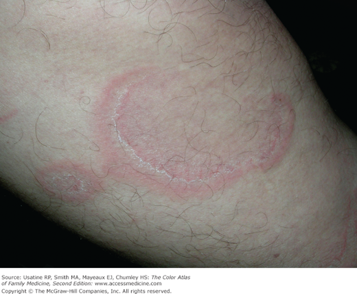
Figure 206-4
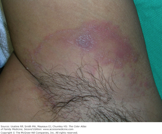
Erythema annular centrifugum in the axilla of a 28-year-old man, which had repeatedly been mistaken for tinea corporis. The trailing scale is visible and a punch biopsy confirmed the diagnosis of erythema annular centrifugum. (Courtesy of Richard P. Usatine, MD
Stay updated, free articles. Join our Telegram channel

Full access? Get Clinical Tree


