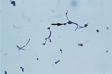
1. Describe the Gram stain morphology of Arcanobacterium, Lactobacillus, Erysipelothrix, and Gardnerella spp. 2. Identify the media of choice and morphologic appearance of Gardnerella sp. and describe its incubation conditions, including time, oxygen requirements, and temperature. 3. List the disease states associated with Erysipelothrix, Gardnerella, and Lactobacillus spp. 4. Identify the correct specimens for the isolation of Erysipelothrix, Gardnerella, and Lactobacillus spp. 5. Explain why in vitro susceptibility testing is usually not necessary to guide therapy of Erysipelothrix or Gardnerella spp. The genera described in this chapter are all catalase-negative, non-spore-forming, gram-positive rods; some may exhibit rudimentary branching. Erysipelothrix rhusiopathiae is one of three species in the genus, but it is considered the only human pathogen. E. rhusiopathiae consists of several serovars based on peptidoglycan structure. The serovars most commonly associated with human infection include serovars 1 and 2. Arcanobacterium spp. demonstrate irregular, gram-positive rods on Gram stain. Gardnerella sp. fermentation byproducts include acetic and lactic acid. The cell wall of Gardnerella sp. is significantly thinner and contains less peptidoglycan than the typically gram-positive bacteria. Weissella confusa, formerly classified as Lactobacillus confusus, is included in Tables 18-3 and 18-4 because it is easily confused on culture media with the organisms included in this chapter, and in rare cases it has been isolated associated with bacteremia and endocarditis. Erysipelothrix spp. are found worldwide in a variety of vertebrate and invertebrate animals, including mammals, birds, and fish. Other domestic animals that may be infected include sheep, rabbits, cattle, and turkeys. The organism may be transmitted through direct contact or ingestion of contaminated water or meat. Arcanobacterium spp. are normal inhabitants of the mucosal membranes of cattle, sheep, dogs, cats, and pigs. The organisms listed in Table 18-1 include those that are closely associated with animals and are contracted by humans through animal exposure (e.g., E. rhusiopathiae and Arcanobacterium pyogenes) and those that are part of the normal human flora (e.g., Lactobacillus spp. and Gardnerella vaginalis). TABLE 18-1 G. vaginalis and Lactobacillus spp. (Table 18-2) are natural inhabitants of the human vagina. Vaginal infections with G. vaginalis are often found in association with a variety of mixed anaerobic flora. Extravaginal infections are uncommon but have been identified associated with postpartum endometritis, septic abortion, and cesarean birth. TABLE 18-2 Pathogenesis and Spectrum of Disease Generally, no special considerations are required for specimen collection and transport of the organisms discussed in this chapter. Of note, skin lesions for Erysipelothrix should be collected by biopsy of the full thickness of skin at the leading edge of the discolored area. Refer to Table 5-1 for other general information on specimen collection and transport. Lactobacillus is highly pleomorphic, occurring in long chaining rods and in coccobacilli and spiral forms (Figure 18-1). Table 18-3 describes the colonial appearance and other distinguishing characteristics (e.g., hemolysis) of each genus on sheep blood agar. G. vaginalis produces small, gray, opaque colonies surrounded by a diffuse zone of beta-hemolysis on HBT agar (Figure 18-2). TABLE 18-3 Colonial Appearance on 5% Sheep Blood Agar and Other Characteristics
Erysipelothrix, Lactobacillus, and Similar Organisms
General Characteristics
Epidemiology
Species
Habitat (Reservoir)
Mode of Transmission
Erysipelothrix rhusiopathiae
Normal flora; carried by and causes disease in animals
Zoonoses; abrasion or puncture wound of skin with animal exposure
Arcanobacterium haemolyticum
Normal flora of human skin and pharynx
Uncertain; infections probably caused by person’s endogenous strains
Arcanobacterium pyogenes
Normal flora; carried by and causes disease in animals.
Uncertain:
Abrasion or undetected wound during exposure to animals
Gardnerella vaginalis
Normal flora: Human vaginal tissue
Colonizers: Distal urethra of males
Endogenous strain
Lactobacillus spp.
Environmental: Widely distributed in foods and nature
Normal flora: Human mouth, gastrointestinal tract, and female genital tract
Endogenous strain
Infections are rare.
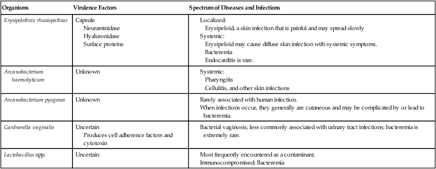
Pathogenesis and Spectrum of Disease
Organisms
Virulence Factors
Spectrum of Diseases and Infections
Erysipelothrix rhusiopathiae
Capsule
Neuraminidase
Hyaluronidase
Surface proteins
Arcanobacterium haemolyticum
Unknown
Arcanobacterium pyogenes
Unknown
Gardnerella vaginalis
Uncertain
Produces cell adherence factors and cytotoxin
Lactobacillus spp.
Uncertain
Laboratory Diagnosis
Specimen Collection and Transport
Direct Detection Methods
Colonial Appearance
Organism
Appearance
Arcanobacterium spp.
Small to large colonies with various appearances, including smooth, mucoid, and white and dry, friable, and gray; may be surrounded by narrow zone of beta-hemolysis
Erysipelothrix rhusiopathiae
Two colony types: large and rough or small, smooth, and translucent; shows alpha-hemolysis after prolonged incubation
Gardnerella vaginalis
Pinpoint; nonhemolytic
Lactobacillus spp.
Multiple colonial morphologies, ranging from pinpoint, alpha-hemolytic colonies resembling streptococci to rough, gray colonies
Weissella confusa
Pinpoint; alpha-hemolytic and may be confused with organisms presented in this chapter. ![]()
Stay updated, free articles. Join our Telegram channel

Full access? Get Clinical Tree


Erysipelothrix, Lactobacillus, and Similar Organisms