Patient Story
A 15-year-old boy is brought in by his mother with a concern about growth of his birthmark. It has become somewhat more raised and bumpy in the past year (Figure 164-1). The adolescent reports no symptoms and is not worried about the appearance. He is otherwise healthy with no neurologic symptoms. The joint decision of the family and the doctor was to not excise the epidermal nevus at this time. He may choose to have this removed by a plastic surgeon in the future.
Introduction
- Epidermal nevi (EN) are congenital hamartomas of ectodermal origin classified on the basis of their main component: sebaceous, apocrine, eccrine, follicular, or keratinocytic.
- Nevus sebaceous (NS) is a hamartoma of the epidermis, hair follicles, and sebaceous and apocrine glands. A hamartoma is the disordered overgrowth of benign tissue in its area of origin.
Synonyms
- EN syndrome is also called Solomon syndrome and is a neurocutaneous disorder characterized by EN and an assortment of neurologic and visceral manifestations.
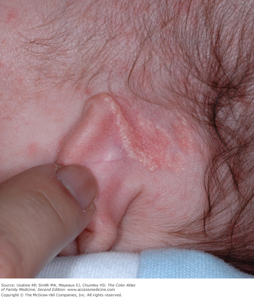
- NS is also called sebaceous nevus and nevus sebaceus of Jadassohn (Figure 164-2).
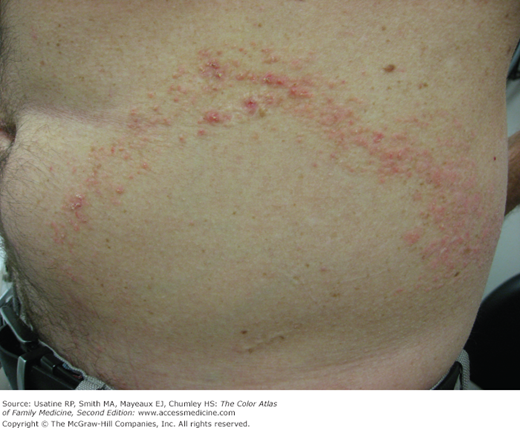
- An inflammatory linear verrucous epidermal nevus (ILVEN) (Figure 164-3) can be part of an epidermal nevus syndrome but some affected persons only have the cutaneous EN.
Epidemiology
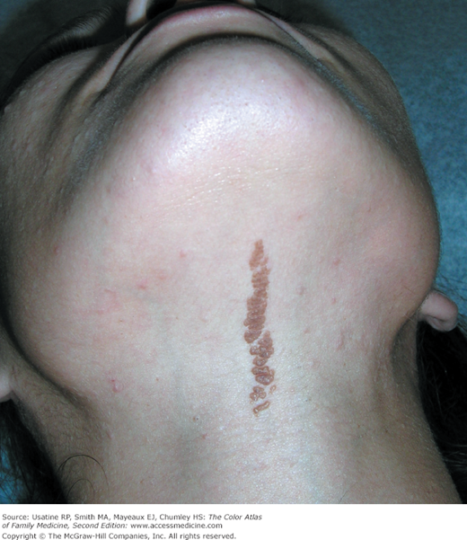
- EN are uncommon (approximately 1% to 3% of newborns and children), sporadic, and usually present at birth, although they can appear in early childhood (Figure 164-4).
- EN are associated with disorders of the eye, nervous, and musculoskeletal systems in 10% to 30% of patients; in one study, 7.9% of patients with EN had 1 of the 9 syndromes—an estimated 1 per 11,928 pediatric patients.1
- In another review of 131 cases of EN, most (60%) had noninflammatory EN, one third had NS, and 6% had ILVEN.2
- NS is usually present at birth or noted in early childhood (Figure 164-5).3 Most cases are sporadic but familial cases have been reported.2
- Linear NS is estimated to occur in 1 per 1000 live births.4
- Linear NS syndrome includes a range of abnormalities, including the central nervous system (CNS); patients with CNS involvement typically have cognitive impairment and seizures3; other organ systems, including the cardiovascular, skeletal, ophthalmologic, and urogenital systems, may be involved.
Etiology and Pathophysiology
- EN histologically display hyperkeratosis and papillomatosis, similar microscopically to seborrheic keratosis (see Chapter 158, Seborrheic Keratosis).2 Also similar to seborrheic keratosis, some EN of keratinocyte differentiation (approximately one third) have been found to have a mutation in the fibroblast growth factor receptor 3 (FGFR3) gene.
- Nine EN syndromes have been reported and are described in the referenced article.5
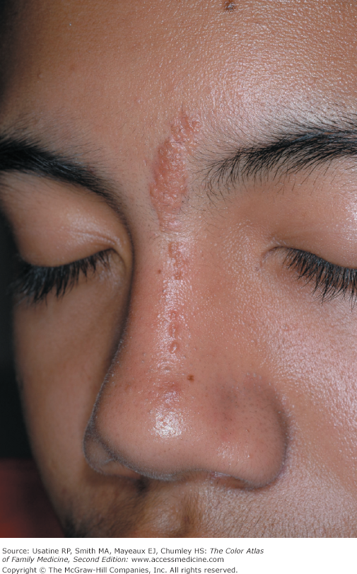
- EN frequently have a linear pattern that follows Blaschko lines (Figures 164-1, 164-3, and 164-4), which are believed to represent epidermal migration during embryogenesis.
- EN tend to become thicker, verrucous (Figure 164-4), and hyperpigmented at puberty.2
- Similarly, NS demonstrates stages of evolution paralleling the histologic differentiation of normal sebaceous gland:6
- Infancy and young children—Smooth to slightly papillated, waxy, hairless thickening (Figure 164-5).
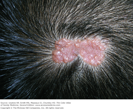
- Puberty—Epidermal hyperplasia resulting in verrucous irregularity of the surface covered with numerous closely aggregated yellow-to-brown papules (Figure 164-6).
- Development of secondary appendageal tumors (Figures 164-7 and 164-8) occurs in 20% to 30% of patients, most are benign (most commonly basal cell epithelioma or trichoblastoma), but single (most commonly basal cell carcinoma) or multiple malignant tumors of both epidermal and adnexal origins may be seen and metastases have been reported.2 Rarely, these malignancies are seen in childhood.
- Infancy and young children—Smooth to slightly papillated, waxy, hairless thickening (Figure 164-5).
- NS was shown to have a high prevalence of human papillomavirus DNA and authors postulate that human papillomavirus (HPV) infection of fetal epidermal stem cells could play a role in the pathogenesis.7


