Localized, wart-like variants, solitary or multiple
• Nevus unius lateris
Long, linear lesions on unilateral extremities
• Ichthyosis hystrix
Large, bilateral lesions on trunk
Etiology/Pathogenesis
• Epidermal nevus syndrome includes neurological, ocular, and skeletal abnormalities
Clinical Issues
• Common sites include neck, trunk, and extremities
• May present together with nevus sebaceus, woolly hair nevus, and nevus comedonicus
• Associated with number of diseases and syndromes; look for other clinical findings
• Small lesion can be excised, larger lesions can be treated by laser or cryotherapy
Microscopic
• At least 10 different patterns, > 1 pattern can coexist in single lesion
• Common pattern includes hyperkeratosis with papillomatosis and acanthosis
• Inflammatory linear verrucous epidermal nevus is considered subtype of epidermal nevus
Top Differential Diagnoses
• Seborrheic keratosis
• Acanthosis nigricans
• Confluent and reticulated papillomatosis of Gougerot and Carteaud
• Organoid nevus (nevus sebaceous)
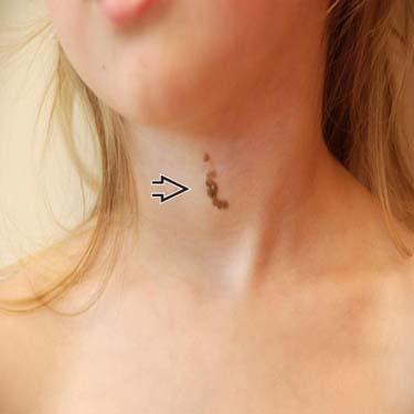
Hyperpigmented Epidermal Nevus An example of an epidermal nevus exhibits hyperpigmented, curvilinear, mammillated plaque on the anterior, midline neck of a girl. (Courtesy J. Finch, MD.)
Classic Histology of Epidermal Nevus Epidermal nevus shows orthokeratotic hyperkeratosis overlying epidermal papillomatosis . Note hyperpigmentation in the basal keratinocytes. (Courtesy C. Cockerell, MD.)
Hypopigmented Epidermal Nevus In this example of epidermal nevus in another child, a cauliflower-like, hypopigmented, and exophytic tumor clinically resembles a wart. Note the finger-like projections . (Courtesy J. Finch, MD.)
Histology Resembles Seborrheic Keratosis Under low magnification, prominent orthokeratotic hyperkeratosis (scale without nucleated cells) and papillomatosis (church spire-like acanthosis) are constant features of epidermal nevus.
TERMINOLOGY
Synonyms
• Nevus verrucosus: Localized, wart-like variants, solitary or multiple
• Nevus unius lateris: Long, linear lesions on unilateral extremities
• Ichthyosis hystrix: Large, bilateral lesions on trunk
Definitions
• Developmental malformation of epidermis with hyperplasia of keratinocytes
• Specific entity that does not include adnexal malformations or appendageal tumors, such as organoid/sebaceous nevus


 on the anterior, midline neck of a girl. (Courtesy J. Finch, MD.)
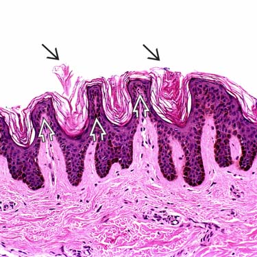
 overlying epidermal papillomatosis
  . Note hyperpigmentation in the basal keratinocytes. (Courtesy C. Cockerell, MD.)
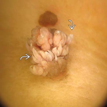
 . (Courtesy J. Finch, MD.)
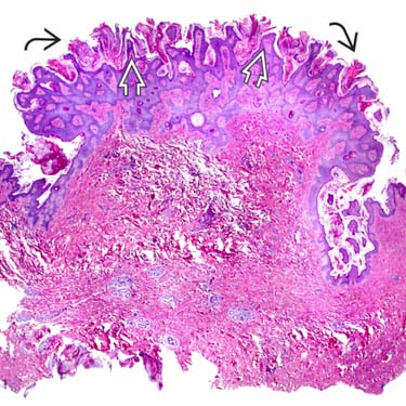
 (scale without nucleated cells) and papillomatosis
  (church spire-like acanthosis) are constant features of epidermal nevus.


