Enchondromatosis
G. Petur Nielsen, MD
Andrew E. Rosenberg, MD
Key Facts
Terminology
Multiple enchondromas that are associated with Ollier disease and Maffucci syndrome
Maffucci syndrome defined as ≥ 3 enchondromas associated with extraosseous hemangiomas
Ollier disease defined as ≥ 3 enchondromas
Clinical Issues
Frequently 1st noted during childhood
Frequently affects short tubular bones of hands
Hemangiomas of Maffucci syndrome usually located in skin or soft tissue
May have anywhere from 3 to hundreds of tumors
Painful lesions need to be biopsied and possibly removed
Patients with Ollier disease or Maffucci syndrome must have lifetime monitoring of their tumors
Image Findings
Tumors are multiple
Some lesions may arise within medullary cavity and resemble solitary enchondroma
Macroscopic Features
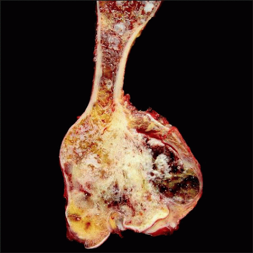
Multiple nodules
Gray glistening in appearance
Microscopic Pathology
Frequently demonstrates increased cellularity and cytologic atypia compared to sporadic enchondromas
Lacks infiltrative growth pattern
May contain myxoid matrix
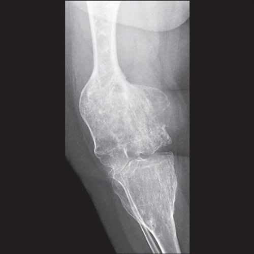
 Radiograph of the right knee shows extensive deformity and shortening of the femur and tibia. The metaphyses of both bones are expanded and contain punctuate cartilaginous calcifications. |
TERMINOLOGY
Definitions
Multiple enchondromas may be associated with Ollier disease or Maffucci syndrome
90% of patients with multiple enchondromas have Ollier disease
10% are associated with Maffucci syndrome
Ollier disease is defined as ≥ 3 enchondromas usually affecting short and long tubular bones
Maffucci syndrome is defined as ≥ 3 enchondromas associated with extraosseous hemangiomas
These hemangiomas were originally classified as cavernous hemangiomas or arteriovenous malformations
More recent studies have shown that most of these vascular tumors are spindle cell hemangiomas
ETIOLOGY/PATHOGENESIS
Neoplastic
Recently, enchondromas in Ollier disease and Maffucci syndrome have been shown to be caused by somatic mutation involving IDH1 and IDH2
CLINICAL ISSUES
Epidemiology
Incidence
Rare; exact incidence is not known
Age
Frequently 1st noted during childhood
In patients with Ollier disease, cartilage tumors may stabilize in size after puberty or continue to grow
Gender
Equal sex distribution
Site
Enchondromas may involve
Only small bones of hands and feet
Only long bones, scapulae, and pelvis
Small, long and flat bones
Involvement of vertebrae and skull is rare
Tends to affect 1 side of body more severely than the other
Within long bones, enchondromas are usually metaphyseal-diaphyseal in location
Hemangiomas in patients with Maffucci syndrome are usually located in skin or soft tissue
May be centered in viscera
1 site of body frequently more severely affected
Presentation
Variable: Frequently, initial symptom is related to enlargement of fingers
Patients may have anywhere from 3 to hundreds of tumors
May be asymptomatic, especially if there are only few enchondromas
May cause severe mechanical and cosmetic problems
Natural History
In patients with Ollier disease, cartilage tumors may stabilize in size after puberty or continue to grow
Development of malignancies within viscera as well as in enchondromas is an important aspect of Maffucci syndrome
Carcinomas of pancreas and malignancies of ovaries, brain, and skeleton are commonplace in Maffucci syndrome
Chondrosarcoma is major malignancy associated with Ollier disease
Treatment
Prognosis
Overall risk of malignant transformation into chondrosarcoma is 40%
More common in patients with severe skeletal involvement or involvement of long and flat bones
IMAGE FINDINGS
Radiographic Findings
Tumors are multiple; some may arise within medullary cavity and resemble solitary enchondroma
Others may be eccentric, arise in cortex or on surfaces of bone, and even span joints
In long bones, persistent columns of dysplastic cartilage that originate in growth plate produce linear radiolucencies that extend from metaphysis into diaphysis
This is pathognomonic for either Ollier disease or Maffucci syndrome
Oblique tubular cartilaginous lesions that follow course of nutrient vessels of bone
Severely affected bones are shortened and deformed
Phleboliths in soft tissue hemangiomas may be present in patients with Maffucci syndrome
Destruction of bone and disappearance of previously seen matrix calcifications point to malignant degeneration
MR Findings
Low signal intensity on T1-weighted images and high signal intensity on T2-weighted images
CT Findings
Enchondromas are well circumscribed and show variable degree of calcifications
Bone Scan
Mild uptake: Demonstrates multiple lesions
MACROSCOPIC FEATURES
General Features
Multiple gray glistening nodules that may coalesce into large masses
MICROSCOPIC PATHOLOGY
Histologic Features
Similar to those of sporadic solitary enchondromas; frequently demonstrate greater degree of cellularity and cytologic atypia and may contain myxoid matrix
DIFFERENTIAL DIAGNOSIS
Chondrosarcoma
In small sample, it can be very difficult to distinguish enchondromas in Ollier disease and Maffucci syndrome from low-grade chondrosarcoma
Most important histologic feature is demonstration of infiltrative growth pattern in chondrosarcoma
SELECTED REFERENCES
1. Pansuriya TC et al: Somatic mosaic IDH1 and IDH2 mutations are associated with enchondroma and spindle cell hemangioma in Ollier disease and Maffucci syndrome. Nat Genet. 43(12):1256-61, 2011
Stay updated, free articles. Join our Telegram channel

Full access? Get Clinical Tree


