Embryonal Rhabdomyosarcoma
Khin Thway, BSc, MBBS, FRCPath
Key Facts
Terminology
Malignant soft tissue tumor that shows variable differentiation toward skeletal muscle
Encompasses botryoid, spindle cell, and anaplastic variants
Clinical Issues
Most common rhabdomyosarcoma (RMS) subtype
Represents 60-70% of RMS
Generally affects younger population than alveolar RMS
Majority in children < 5 years
Head and neck
Genitourinary region
Other sites include retroperitoneum, pelvis, and biliary tract
Microscopic Pathology
Loose fascicles and sheets
Cytology, pattern, and cellularity can vary
Spindle, stellate, and ovoid cells
Rhabdomyoblasts in variable numbers and stages of differentiation
Botryoid RMS
Grows beneath epithelial surfaces
Spindle cell RMS
Spindle cells with elongated nuclei
May exhibit cross-striations
Anaplastic RMS
Atypical or bizarre tumor cells, present focally or more diffusely
Complex karyotypes
Associated with 11p15.5 loss of heterozygosity
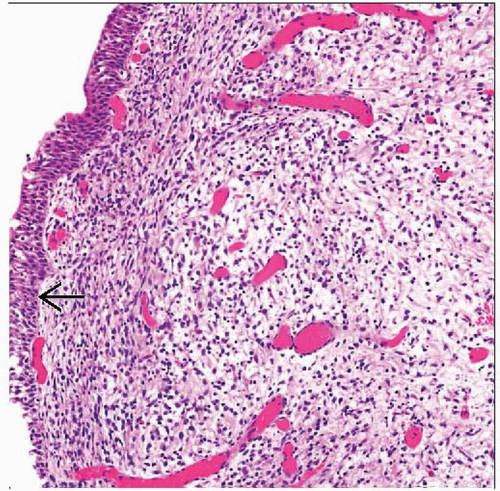 Embryonal rhabdomyosarcoma of the bladder is seen under urothelium  . It is mostly of low cellularity and not easily identifiable at low power when the appearances can be mistaken for inflammation. . It is mostly of low cellularity and not easily identifiable at low power when the appearances can be mistaken for inflammation. |
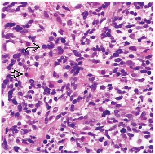 The tumor cells show a range of appearances. There are relatively undifferentiated ovoid cells  along with rhabdomyoblasts with prominent eosinophilic cytoplasm and eccentric nuclei along with rhabdomyoblasts with prominent eosinophilic cytoplasm and eccentric nuclei  . . |
TERMINOLOGY
Abbreviations
Embryonal rhabdomyosarcoma (ERMS)
Definitions
Malignant soft tissue tumor that shows variable differentiation toward embryonic skeletal muscle
ERMS encompasses botryoid, spindle cell, and anaplastic variants
ETIOLOGY/PATHOGENESIS
Unknown
Cell of origin still unknown
Possible candidate cells include muscle stem cells and multipotent mesenchymal stem cells
Often occur in sites lacking skeletal muscle
e.g., bladder, prostate
CLINICAL ISSUES
Epidemiology
Incidence
Rhabdomyosarcomas (RMS) are most frequent soft tissue sarcomas in children and young adults
ERMS is most common RMS subtype
4.3 cases per 1,000,000 children/year in USA
Represent 60-70% of RMS
Age
Children
ERMS generally affects younger population than alveolar RMS
Typically < 10 years of age
Majority in children < 5 years
Bimodal distribution with smaller peak in adolescence
Occasional cases congenital
Rarer in adults
Where pleomorphic subtype predominates
Gender
Slight male preponderance
M:F= 1.4:1
Site
Head and neck
Particularly orbital and parameningeal sites
Genitourinary region
Bladder
Prostate
Paratesticular soft tissue
Other sites
Retroperitoneum and pelvis
Biliary tract
Much less frequent involvement of trunk and limbs than alveolar rhabdomyosarcoma (ARMS)
Spindle cell RMS
Paratesticular region
Botryoid RMS
Arise beneath mucosal epithelial surfaces
Bladder
Vagina
Extrahepatic bile ducts
More rarely, auditory canal or conjunctiva
Presentation
Suddenly enlarging mass
Local symptoms pertaining to site of origin
e.g., deafness, proptosis (head and neck)
e.g., urinary retention (genitourinary sites)
Prognosis
Main prognostic parameters are histologic type, disease stage, and site
Favorable sites are head and neck (nonparameningeal), genitourinary (nonbladder, nonprostate), and bile duct
Botryoid and spindle cell variants (excluding aggressive adult spindle cell variant) have better prognosis
ERMS has significantly better prognosis than ARMS
5-year overall survival approximately 73%
MACROSCOPIC FEATURES
General Features
Fleshy mass
Margins usually infiltrative
Tan to white
Rubbery cut surface
Hemorrhage
Necrosis
Cystic degeneration
Botryoid RMS
Exophytic, polypoid tumor
More circumscribed margins
Small nodules adjacent to mucosal surface
Masses may fill lumen of hollow viscus
Gelatinous cut surface
MICROSCOPIC PATHOLOGY
Histologic Features
Patterns variable
Loose fascicles and sheets
Variable cellularity
Can alternate between markedly cellular and looser myxoid zones
Spindle, stellate, and ovoid cells
In varying stages of myogenic differentiation
Ovoid and elongated nuclei
Nuclei hyperchromatic or vesicular
Rhabdomyoblasts
Cells with eccentric nuclei and variable amounts of eosinophilic cytoplasm
Cytoplasmic cross-striations may be visible
Variable numbers and stages of differentiation
Varying shapes
Strap cells
Tadpole cells
Spider cells
Myxoid stroma
Mitoses usually easily discernible
Necrosis
Tumor giant cells are rare, in contrast to alveolar RMS
Botryoid RMS
Polypoid
Cambium layer
Tightly packed cellular layer of tumor cells closely abutting epithelial surface
Loose myxoid stroma
Can be of relatively low cellularity
May be missed or mistaken for chronic inflammation
Spindle cell RMS
Spindle cells with elongated nuclei
May exhibit cross-striations
Tumors may have abundant collagenous stroma
Anaplastic RMS
Pleomorphic cells
Atypical or bizarre tumor cells
Anaplasia may be present as scattered cells or as foci or large sheets of cells
Presence of anaplastic cells in aggregates or diffuse sheets associated with poorer survival
Atypical mitotic figures
Stay updated, free articles. Join our Telegram channel

Full access? Get Clinical Tree


