Effusions in the Presence of Cancer


Effusions in the Presence of Cancer


Cytologic techniques have been universally recognized as the most important diagnostic tool in the recognition of malignant tumors in effusions. The diagnosis of cancer in
a pleural, pericardial, or peritoneal fluid is of capital importance for the patient and the attending physician or surgeon. Although, in many such instances, a fatal outcome of the disease may be anticipated, some tumors offer a much better prognosis than others. For example, metastatic mammary carcinoma may be controlled, often for a period of many years, by various forms of therapy. In some other tumors, long-term remissions or even cures can be achieved. Malignant lymphomas, some testicular tumors, and some malignant tumors of childhood, such as neuroblastoma or embryonal rhabdomyosarcoma, may respond to energetic therapeutic measures. Therefore, the responsibility of the pathologist is two-fold:


  • To identify cancer cells accurately


  • To identify tumor type and, if possible, the site of primary origin

These tasks are greatly facilitated by an accurate clinical history and review of prior histologic material, if available.

As a general rule, it is better to exercise diagnostic caution, keeping in mind the potentially tragic consequences of an erroneous diagnosis of cancer based on flimsy evidence. On the other hand, failure to recognize cancer cells that show only subtle morphologic abnormalities may delay or deprive the patient of needed treatment.

The use of impeccable technical preparations is of utmost importance in ensuring diagnostic accuracy. The collection techniques are discussed in Chapter 1 and the laboratory processing techniques are discussed in detail in Chapter 44. Preparation of cell blocks from residual sediment is often of great diagnostic value in the recognition of morphology and origin of the tumor and in the application of special stains or other analytical procedures.

Experimental studies by Siegler and Koprowska (1962) on the mechanism of ascites formation in mice indicated that the formation of ascites, containing malignant cells, was conditioned by damage to the capillaries and lymphatics by colonies of cancer cells. It is likely that a similar mechanism is operative in humans.


RECOGNITION OF MALIGNANT CELLS

The frequently emphasized difficulty in the recognition and classification of cancer cells in body fluids is caused by two main factors:


  • The body fluids are a natural tissue culture medium, wherein mesothelial and tumor cells may proliferate free of the boundaries imposed upon them by the framework of organs and tissues (also see comments in Chap. 25). It is known to all students of in vitro tissue culture that morphologic identification of benign versus malignant cultured cells may be fraught with considerable difficulty. Similarly, the characteristic features of human cancer cells in fluids may undergo substantial modifications. For example, abnormal cell shapes that often help in the identification of exfoliated or aspirated cancer cells may no longer be present in fluids, wherein the cancer cells may assume a neutral, spherical appearance. Nuclear features often seen in cancer, such as hyperchromasia, may also be absent or attenuated.


  • Proliferating mesothelial cells may conceal the presence of tumor cells or may mimic cancer cells by forming complex clusters or displaying alarming nuclear features, such as the presence of nucleoli (see Chap. 25). Rarely, clusters of macrophages (histiocytes) may mimic cancer.

In spite of these words of caution, it is entirely possible, in the vast majority of effusions, to identify cancer cells accurately, often to identify tumor type, and, sometimes, to suggest the primary tumor of origin, even in the absence of an accurate clinical history. The diagnostic value and significance of ancillary diagnostic procedures are discussed below (Table 26-1).

The general features leading to the recognition of cancer cells in smears and similar preparations are described at length in Chapter 7. The loss of some of these features in effusions requires close attention to morphologic details that may be of secondary value in other diagnostic media.


Cytoplasmic Features


Cell Size

The size of tumor cells may vary greatly according to tumor type. To determine the size of a suspect cell, a comparison must be made with identifiable cell types, such as erythrocytes, lymphocytes, or mesothelial cells. Generally, the cells of malignant tumors in effusions may be classed in three size groups:


  • Large or very large. The cells are significantly larger than normal mesothelial cells. Some mesotheliomas, metastatic carcinomas of various types, malignant melanomas and sarcomas belong in this group. When combined with abnormal nuclear features, described below, the identification of such tumors is easy (see Figs. 26-36B, 26-40D, 26-57).


  • Small. The tumors are made up of cells much smaller than mesothelial cells. Most malignant lymphomas, many of the malignant tumors of childhood (neuroblastoma, Wilms’ tumor) and certain carcinomas (small-cell carcinoma of the breast, oat cell carcinoma) belong to this group of tumors. Close attention must be paid to nuclear features and interrelationship of cells for accurate identification (see Figs. 26-28, 26-30, 26-35).


  • Medium-sized. The diagnostic problem usually occurs with cells of medium size which are approximately of the same size as mesothelial cells. A variety of carcinomas of mammary, lung, gastric, pancreatic, or prostatic origin may have this presentation (see Fig. 26-26). This is perhaps the most difficult group of tumors to identify and the most important source of diagnostic error.


Cell Configuration

Because many cancer cells in fluids assume a “neutral” spherical configuration, unusual cell shapes are very helpful
in identifying cancer. Thus, the presence of bizarre or spindly cells (see Figs. 26-19, 26-20, and 26-57), columnar cells or cells resembling bronchial lining cells is usually associated with cancer (see Figs. 26-34, 26-41). Exceptions may occur, for example in the sediment in rheumatoid arthritis wherein spindly epithelioid cells may be observed (see Chap. 25). Very rarely, benign fibroblasts, derived from connective tissue supporting the mesothelium, may also be observed in such fluids.


TABLE 26-1 CELL FEATURES USEFUL IN THE IDENTIFICATION OF CANCER CELLS IN EFFUSIONS


Cell size


Diagnostic value


Benign sources of error


Small


Oat cell carcinoma, some breast cancers, small cell lymphomas, tumors of childhood


Chronic lymphocytic infiltrate (e.g., tuberculosis)


Medium


Many carcinomas


Atypical mesothelial cells


Large


Soft-part sarcomas, some carcinomas


Reactive giant cells (Langhans’)


Cell configuration


Spherical


Dominant in carcinomas


Bizarre or spindly


Usually diagnostic of a malignant tumor


Rheumatoid arthritis, benign fibroblasts


Other cytoplasmic features


Keratin formation


Diagnostic of squamous cancer


Squamous epithelial cells from skin (vanishingly rare)


Mucus formation


Diagnostic of metastatic carcinomas of various derivations


No benign counterpart


Cell products


Melanin pigment


Nearly always diagnostic of malignant melanoma. For exceptions, see text.


Hemosiderin and other hemoglobin-derived pigments


Psammoma bodies


Ovarian, thyroid, mesotheliomas, other rare carcinomas


Dystrophic calcifications of serous cavities (exceptional)


Cell surfaces


Long microvilli (light and electron microscopy)


Mesotheliomas and many carcinomas (not oat cell)


Short orderly surface microvilli on mesothelial cells (on electron microscopy only)


Cell aggregates


Numerous large three-dimensional with lumens on cross section (cell blocks)


Carcinomatous mesothelioma; many carcinomas


Mesothelial cell aggregates, usually small and flat.


Exceptionally, macrophages (histiocytes) in nodular histiocytic hyperplasia


Nuclear features


Less hyperchromatic than in other diagnostic media


Many malignant tumors


Rarely enlarged nucleoli in mesothelial cells


Abnormal N/C ratio


Large nucleoli


Very rare in mesothelial cells


Bizarre nuclear shapes (protrusions) and massive apoptosis (karyorrhexis)


Malignant lymphomas


No benign counterpart


Intranuclear cytoplasmic inclusions (nuclear “holes”)


Carcinoma of thyroid, melanoma


Mitoses


Normal configuration


Suspicious of cancer


Rare normal and exceptionally abnormal mitoses of mesothelial cells and macrophages


Abnormal configuration


Diagnostic of cancer


Other Cytoplasmic Features

Besides cell size and configuration, there are a few other cytoplasmic features that are sometimes helpful in the identification of cancer cells. Keratin formation, expressed as thick cytoplasm, staining orange or yellow in Papanicolaou stain, is practically synonymous with squamous cancers (see Figs. 26-23 and 26-32). Large, mucus-containing vacuoles, if accompanied by nuclear abnormalities, occur in cells of metastatic adenocarcinomas of various derivations
(see Fig. 26-41). Such cells must be differentiated from “signet ring” macrophages that have normal, small nuclei (see Chap. 25). Intracytoplasmic glandular inclusions (“target cells” or “bull’s eye cells”) are observed mainly in mammary carcinoma (see Fig. 26-28D) but may sometimes occur in other tumors as well (Kumar et al, 2000).

Rarely, other cytoplasmic features may assist in the identification of tumor types. For example, cancer cells derived from striated muscle, may show the presence of cytoplasmic cross-striations (see Fig. 17-6). Such cells are diagnostic of metastatic rhabdomyosarcomas or tumors with a rhabdomyosarcoma component, such as metastatic mesodermal mixed tumors.


Cell Products

Products of metabolic activity of cells, such as mucus (demonstrated by mucicarmine stain), in cytoplasmic vacuoles, is very rarely, if ever, produced by benign cells in effusions. Accumulation of intracytoplasmic melanin pigment (not to be confused with other pigments; see Chap. 25) is nearly always diagnostic of malignant melanoma or related tumors (see Fig. 26-56). Calcified, concentrically laminated, round or oval psammoma bodies, 20 to 50 μm in size, are most commonly observed in metastatic tumors of ovarian origin (see Fig. 26-36), but may also be produced by thyroid cancer (see Fig. 26-43), carcinomatous mesotheliomas (see Fig. 26-17) and, very rarely, by bronchogenic adenocarcinoma, pancreatic carcinoma, carcinoma of the renal pelvis, endometrial carcinoma, and mammary carcinoma. It must be noted that the presence of calcified bodies is of limited diagnostic value in cul-de-sac pelvic washings (see Chap. 16).


Cell Surfaces

Spriggs and Meek (1961) observed the presence of tufts of hair-like processes on the surfaces of some malignant cells in pleural and peritoneal effusions, sometimes limited to one segment, but often covering the entire cell (Fig. 26-1). The processes were particularly striking in certain cases of metastatic ovarian carcinoma. Ebner and Schneider (1956) also reported the presence of “ciliated malignant cells” in carcinoma of the ovary.

These early studies have been extended by Domagala and Woyke (1975) and, subsequently, Domagala and Koss (1977). With scanning electron microscopy, it was shown that the surfaces of most cancer cells in effusions, regardless of tumor type or origin, were covered by innumerable microvilli of variable shape and configuration (anisovillosis) (Fig. 26-2). A notable exception were oat carcinoma cells that had a surface without microvilli. The scanning electron microscopic appearance of the surfaces of cancer cells was markedly different from surface configuration of macrophages and mesothelial cells, described in Chapter 25. Subsequent studies have shown that cells of carcinomatous mesothelioma had particularly long, complex microvilli on their surfaces, a feature that is sometimes of diagnostic value (see Fig. 26-1). The significance of microvilli is not understood at the time of this writing (2004).


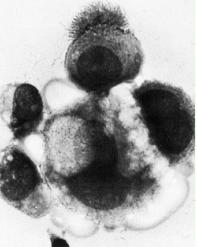
Figure 26-1 Cluster of cells in a malignant mesothelioma of tunica vaginalis testis in a 21-year-old man. A tuft of long microvilli may be observed on one of the cells. (× 2,000.) (Courtesy of Dr. Arthur Spriggs, Oxford, England)

The microvilli can be observed in light microscopy in the form of a striated halo, particularly on surfaces of air-dried cancer cells (Fig. 26-3). The presence of visible surface microvilli may be helpful in the recognition of malignant cells.


Cell Aggregates

Although benign mesothelial cells may form aggregates in effusions, as described in Chapter 25, the aggregates are usually few, and are usually made up of a small number of cells, rarely more than 20 or 25. Further, the aggregates of benign cells are usually arranged in a monolayer. Exceptions to this rule have been discussed in Chapter 25. When mesothelial cell aggregates are numerous and composed of a large number of cells, they mimic aggregates formed by malignant tumor cells.

Many malignant tumors, principally adenocarcinomas of various primary origin, form cell aggregates, often composed of a very large number of cells. Such aggregates are usually three-dimensional, i.e., made up of several superimposed cell layers that cannot be brought into a single focus. Approximately round or spherical aggregates, corresponding to papillary projections, or aggregates forming gland-like structures with a central lumen (Fig. 26-4A,B), are particularly helpful in identifying adenocarcinomas. In cross-sections of these aggregates in cell blocks, a central lumen is often observed, documenting the glandular or tubular nature of these clusters (Fig. 26-4C,D). The name of “spheroids” or “hollow spheres” has been used by some observers to describe such clusters.

Spriggs (1984), in a detailed light and electron microscopic study of cell aggregates in effusions, pointed out that
these commonly encountered cell clusters are actually elaborate, organized 3-dimensional structures. Spriggs divided the structures into three groups: papillary, tubulopapillary, and acinar (Fig. 26-5). On cross-section, the cells composing the clusters surround a lumen, form cell junctions, and often contain central deposits of collagen as supporting structures. The papillary and tubulopapillary clusters are provided with microvilli on their outer surfaces, whereas the acinar structures contain microvilli on the surface facing the lumen. Thus, the cell aggregates, far from being haphazard accumulations of epithelial cells, are, in fact, highly organized structures formed by benign or malignant cells, growing freely in effusions.


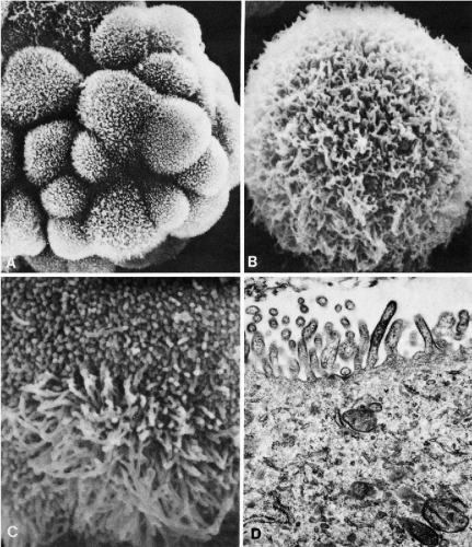
Figure 26-2 Scanning and transmission electron microscopy (TEM) of cancer cells in effusions. A. Cluster of cancer cells from a metastatic ovarian carcinoma. Note innumerable microvilli on the surfaces of cancer cells. B. Breast cancer cell. Note shaggy appearance due to numerous slender microvilli. C. Lung cancer cell (adenocarcinoma). Short stubby microvilli are adjacent to long slender microvilli. D. Cell of ovarian carcinoma in TEM. Note innumerable microvilli of uneven size and configuration on the cell surface. (A: × 2,300; B: × 4,600; C: × 6,000; D: × 18,600.) (Courtesy of Dr. W. Domagala, formerly of Montefiore Hospital, New York, NY.)


Nuclear Features


Nuclear Configuration, Size, and Shape

As has been mentioned above, nuclear hyperchromasia and coarse granularity of chromatin, important landmarks of cancer cells in other media, may not be evident in effusions. The nuclei may be homogeneous and opaque and sometimes clear and transparent.


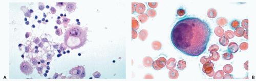
Figure 26-3 Microvilli on the surface of cancer cells. A. Alcohol fixation. B. Air-dried smear (oil immersion). The microvilli are better seen in the air-dried material.

Still, most malignant cells in fluids have enlarged nuclei, corresponding to increased DNA content. In most but not all carcinomas, the nucleocytoplasmic ratio is modified in favor of the nucleus, particularly when compared with mesothelial cells (see Fig. 26-36). However, in some mucus-producing and keratinizing squamous cancer cells, the cytoplasm is abundant and the nucleocytoplasmic ratio may not be conspicuously abnormal.


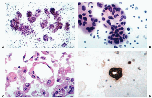
Figure 26-4 Spherical clusters characteristic of malignant mesothelioma and adenocarcinoma. A. A large number of spherical clusters in pleural fluid in a 43-year-old man with malignant mesothelioma. B. High-power view of one of the clusters from A showing a spherical configuration of the cells forming the cluster. C. Cross-section of one of the spherical clusters showing formation of an empty (gland-like) space within the cluster. D. A cell block from another case showing the glandular arrangement of the cells of adenocarcinoma immunostained for low density keratin.

The nuclear shapes of cancer cells in fluids are rarely abnormal. Most such cells display spherical or oval nuclei
with smooth borders. Occasionally, on closer scrutiny, an irregular nuclear outline may be observed and is of diagnostic assistance, particularly in malignant lymphomas, in the form of indentations or protrusions of the nuclear membrane. Another nuclear feature observed in malignant effusions, particularly in malignant lymphomas, is massive nuclear breakdown (apoptosis or karyorrhexis) that is virtually never seen in benign fluids (see Figs. 26-46, 26-47).


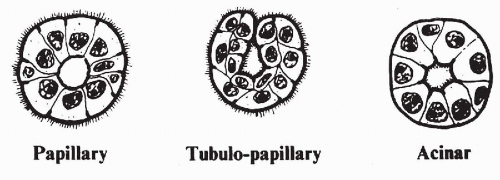
Figure 26-5 Spriggs’ representation of cell clusters in effusions. The clusters are organized structures that may be either papillary with microvilli on the outer surface; tubulopapillary with microvilli on both surfaces; or acinar, with microvilli on the inner luminal aspect of the cluster. (Spriggs AI. The architecture of tumor cell clusters in serous effusions. In Koss LG, Coleman DV (eds). Advances in Clinical Cytology, Vol. 2. New York, Masson, 1984.)


Nucleoli

The presence of nucleoli is of capital diagnostic importance in the recognition of cancer cells in effusions. Except in keratinizing squamous carcinomas, large, irregularly shaped, single or multiple nucleoli are frequently observed in cancer cells (see Fig. 26-55). As described in Chapter 25, on rare occasions, enlarged nucleoli may occur in mesothelial cells, which then become an important source of error.


Figure 26-6 Abnormal mitoses in cancer cells in fluids. A. Quadripolar mitosis in pericardial fluid with metastatic carcinoma. B. An abnormal metaphase in pleural fluid in the presence of a sarcoma. C. Abnormal mitosis in pleural fluid in metastatic carcinoma. D. Typical prophase in the nucleus of a cancer cell in pleural fluid (A,B,D: high magnification.)


Mitoses

Mitoses are very rare in benign fluids and, therefore, their presence should be considered as presumptive evidence of cancer. Abnormal mitotic figures, such as an increase in the number of chromosomes, multipolar mitoses or chromosomal lag (see Chap. 7), are one of the most reliable identifiers of cancer cells in effusions (Fig. 26-6A-C). Occasionally, the nuclear area is filled with
chromatin granules, suggestive of a mitotic prophase (Fig. 26-6D). These findings are uncommon in benign cells and should also be considered as a possible presumptive evidence of a malignant tumor.

However, as discussed in Chapter 25, we observed morphologically atypical mitoses in mesothelial cells in pleural fluid in the absence of cancer in two patients, out of many thousands examined. Papanicolaou, in his Atlas (1954), also mentioned two similar cases. It may be stated safely that such occurrences are extraordinarily rare and should not detract from the diagnostic value of abnormal mitotic figures, which very strongly suggest a malignant process. It is conceivable that chromosomal abnormalities observed in mesothelial cells in ascites in cirrhosis of the liver, discussed in Chapter 25, may account for the rare mitotic abnormalities observed in benign effusions.


Multiple Sex Chromatin (Barr) Bodies

As mentioned in Chapters 7 and 11, in female patients, two or more sex chromatin bodies in the same nucleus are virtually diagnostic of cancer because they document the presence of an abnormal chromosomal complement. This observation is particularly helpful in the diagnosis of some cases of metastatic mammary carcinoma, wherein the size of the cancer cells may be comparable to that of mesothelial cells or macrophages, and the morphologic abnormalities are not pronounced (see Fig. 26-27B). Mesothelial cells and macrophages (histiocytes) have a single sex chromatin body, except in the extraordinary rare superfemales (karyotype 47, XXX). The multiplicity of sex chromatin bodies in cancer cells is sometimes difficult to ascertain because large chromocenters, located near the nuclear membrane, may be misinterpreted as Barr bodies. Thus, the shape and location of the sex chromatin body must be carefully assessed (for further discussion and description of sex chromatin bodies, see Chaps. 4, 7, and 21).


Nuclear Cytoplasmic Inclusions (Orphan Annie Nuclei)

Sharply demarcated clear areas within the nucleus, corresponding to cytoplasmic invaginations, have been observed in a variety of cancer cells, but mainly in cells of metastatic melanomas, thyroid cancers and pulmonary adenocarcinomas (see Fig. 26-55). We have never observed this feature in benign cells in effusions.


ANCILLARY TECHNIQUES IN THE RECOGNITION OF CANCER CELLS IN EFFUSIONS


Cytogenetics

The finding of cells with abnormal chromosomal numbers and configuration has been shown to be diagnostic of cancer cells in effusions (Goodlin, 1961; Ishihara and Sandberg, 1963; Jackson, 1967).

Benedict et al (1971) pointed out that a long acrocentric chromosome was often associated with metastatic malignant tumors, regardless of primary origin and histologic type. Miles and Wolinska (1973) compared the sensitivity of cytogenetic studies with light microscopic diagnoses in 58 cancer patients. In 38 patients, routine cytology disclosed cancer, whereas cytogenetic studies were positive in only 24 of these patients. However, in two patients, chromosome analysis disclosed an aneuploid chromosomal component, whereas routine cytology was negative. Cytogenetic studies in this group of patients may have been handicapped by prior treatment. In general, tissue culture technique is used to obtain a sufficient number of metaphases for cytogenetic analysis. Otherwise, a very large population of dividing cancer cells is required for a successful direct chromosomal analysis.

The apparent exceptions to this rule are the observations by To et al (1981) and by Watts et al (1983) who observed abnormal chromosomal components in several ascitic fluids associated with liver cirrhosis and in one pleural effusion associated with pneumonia (see Chap. 25). Apparently, other disorders, such as rheumatoid arthritis or pulmonary embolus, may occasionally be associated with chromosomal abnormalities (summary in Watts et al, 1983). These studies did not include chromosomal banding. It would be of great interest to confirm these observations by contemporary cytogenetic techniques.

With the introduction of molecular probes to various chromosomes, it became possible to determine chromosomal abnormalities by the technique of fluorescent in situ hybridization (FISH) in interphase nuclei. The principles of this technique are discussed in Chapter 4. The application of this technique to malignant mesotheliomas in effusions is discussed below (Granados et al, 1994). Florentine et al (1997) used probes to chromosomes 3, 8, 10, and 12 to determine numerical chromosomal aberrations, and compared the results of chromosomal analysis with conventional cytology on ThinPrep (Cytyc Corporation, Boxborough, MA) slides of effusions with mixed results. Cajulis et al (1997) specifically identified difficult-to-classify “atypical” cells in previously stained smears in a variety of samples (including four effusions). They used the FISH technique to determine numerical aberrations of chromosome 8. Cajulis observed chromosomal aberrations in most “atypical” cells but not in benign cells and considered the FISH technique with chromosome 8 to have a specificity of 100% and sensitivity of 83%. The reader is cautioned that the FISH technique requires a dedicated laboratory and that the molecular probes to individual chromosomes or their centromeres are expensive. Nonetheless, the results cited are most encouraging and suggest that the technique may prove to be very useful in determining the presence or absence of cancer in difficult cases. The results of FISH technique in cells in the urinary sediment are discussed in Chapter 23.

A novel approach to the identification of cancer cells is the documentation of telomerase activity. Telomeres are the structures capping the ends of normal chromosomes. With each cell division, the telomeres become shorter, resulting in cell senescence. It is assumed that the enzyme telomerase is capable of synthesis of telomeres, thus conferring
immortality on cancer cells. Telomerase activity can be demonstrated by molecular biologic techniques or by immunochemistry using an in situ fluorescent assay and a telomere repeat amplification protocol (TRAP) (Ohyashiki et al, 1997). The test results in nuclear fluorescence in cancer cells whereas, in benign cells, the fluorescence is limited to the cytoplasm. Dejmek et al (2001) adopted this technique to cells in 16 effusions and claimed that the test was specific for cancer cells. Similar data were previously provided for cancer cells in the respiratory tract (Dejmek et al, 2000) and in urinary sediment (Ohayashiki et al, 1998). However, Braunschweig et al (2001) denied any diagnostic value to the TRAP reaction because of frequent false-positive and false-negative results.


DNA Measurements in Effusions as Tumor Markers

Freni et al (1971) and Krivinkova et al (1976) were apparently the first groups of investigators to recognize the diagnostic value of DNA measurements by cytophotometry in the identification of malignant cells in effusions. The presence of cells with abnormal DNA content was well correlated with the presence of cancer.

With the introduction of flow cytometry, the DNA measurements became more rapid and several groups of investigators reported abnormal DNA histograms in fluids containing malignant cells (Evans et al, 1983; Unger et al, 1983; Katz et al, 1985; Croonen et al, 1988). In a study from this laboratory, Schneller et al (1987) pointed out that static cytophotometry may disclose abnormal DNA values in cancer cells that are not revealed by flow cytometry. Further, some malignant tumors are diploid and have a perfectly normal DNA ploidy that cannot be detected by any measurements. Agarwal et al (1991) observed that some benign tumors have an aneuploid DNA distribution. Thus, an abnormal DNA histogram usually (but not always) indicates the presence of cancer but a normal diploid histogram does not necessarily rule out cancer. For further discussion of cytophotometry and flow cytometry, see Chapters 46 and 47.


Cytochemistry and Immunocytochemistry

Cells in effusions are the favored target of cytochemical and immunocytochemical investigations because of abundant cell populations and the ease with which multiple samples can be obtained in the form of smears, cytocentrifuge preparations (cytospins), cell blocks (cell buttons) or the newer methods of processing of liquid samples (ThinPrep). There are no specific cytochemical or immunocytochemical reagents that could distinguish benign from malignant cells. The best effort along these lines was the use of an antibody to the mutated p53 molecule that is commonly expressed in human malignant tumors and practically never in normal tissues (see Chaps. 3 and 7). Mullick et al (1996) applied this antibody to 103 effusions and reported positive staining in 55% of malignant tumors and none in benign controls. Otherwise, these techniques are sometimes helpful in distinguishing from each other tumors of diverse origins and type.

The most useful cytochemical stains are mucicarmine that are frequently helpful in differentiating cancer cells from mesothelial cells, stains for the identification of pigments, such as melanin and some silver stains, all discussed in Chapter 44. The number of monoclonal antibodies tested on effusions is very large and the principal observations are discussed in Chapter 45. Hence, only a brief summary of the most useful antibodies is shown in Table 26-2. A multiple-well technique, which permits synchronous testing of several aliquots of cells with several monoclonal antibodies, was described by Guzman et al (1988).

Regardless of results, the immunocytochemical observations must be considered a secondary mode of cancer cell identification that may sometimes enhance, but never replace, morphologic observations.


Immunologic Response

Another approach of current interest in the study of effusions is the relationship of various cell populations engaged in immune responses to cancer cells in effusions.

Scanning electron microscopic studies by Domagala and Koss (1977) strongly suggested that cell contacts between lymphocytes and macrophages and between macrophages and cancer cells may occur in effusions (Fig. 26-7A). The latter relationship has since been confirmed in light microscopy. Cancer cells in contact with variable numbers of macrophages have been repeatedly observed (Fig. 26-7B). Phagocytosis of cancer cells, either by macrophages or by other cancer cells may also be observed (Fig. 26-7C).

Domagala et al (1978, 1981) also studied the distribution of B and T lymphocytes in the peripheral blood and in effusions of patients with metastatic carcinoma of various primary origins. In most cases, there was a statistically significant increase of T lymphocytes in fluids with metastatic cancer. Similar observations have been made by Djeu et al (1976). Green and Griffin (1996) observed an increase in the subset of lymphocytes known as natural killer cells (identified by antibodies to CD16/CD56) in 14 of 15 patients with metastatic carcinomas in pleural effusions. However, mesotheliomas, lymphomas and leukemias did not show this abnormality. These observations were confirmed by Laurini et al (2000) who used flow cytometry with the same monoclonal antibodies in their studies. These reports suggest that immune mechanisms are operative in some effusions with metastatic cancer and that their further exploration may be of diagnostic and perhaps prognostic significance.


IDENTIFICATION OF TUMOR TYPES

The diagnosis of tumor types, such as adenocarcinomas, squamous carcinomas, tumors with endocrine function, malignant lymphomas, or sarcomas in effusions, is of
significant clinical value. This information may help in the determination of the organ of origin of the tumor and provide guidance to optimal treatment. The best chances of identification of tumor type occur when cancer cells form multicellular structures akin to those observed in tissues or if fragments of tumor can be recognized in cell blocks. Other identification options are based on cell relationships and identification of cell products. The three most common types of tumors encountered in effusions are: adenocarcinomas, poorly differentiated carcinomas of various origins, and small cell tumors. Less often, keratinizing squamous carcinoma may also be recognized. Many of the features of malignant cells described above may serve to identify tumor types.


TABLE 26-2 MONOCLONAL ANTIBODIES USED ON EFFUSIONS


Antibody


Purpose


Keratin: AE1/AE3


To distinguish carcinoma from other types of tumors.


Common lymphocyte antigen: LCA


To mark lymphoid cells.


Lymphocyte markers: CD3


In combination with CD 20, to distinguish lymphoma from reactive lymph node.


Lymphocyte markers: CD20


In combination with CD 3, to distinguish lymphoma from reactive lymph node.


Endocrine markers: Synaphophysin


For neuroendocrine tumors.


Endocrine markers: Chromogranin


For neuroendocrine tumors.


Calretinin


To distinguish mesothelial cells from other epithelial cells.


HMB 45


To favor melanoma.


Intermediate filament: Vimentin


For mesenchymal lesion, and in combination with AE1/AE3 for renal cell carcinoma.


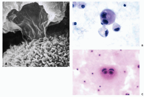
Figure 26-7 Macrophages and phagocytosis in cancer. A. Scanning electron micrograph showing an extension of the cytoplasm of a macrophage onto the surface of a cancer cell identified by microvilli. B. High magnification view of a large cancer cell surrounded by macrophages, corresponding to the scanning electron microscopy image shown in A. C. Phagocytosis of cancer cells, presumably by other cancer cells. This cell arrangement is not uncommon in cancer but may also occur in cirrhosis of the liver.


Adenocarcinomas

Adenocarcinomas of various origins are by far the most common type of tumors encountered in effusions. The common features of adenocarcinomas in fluids are:


Jun 8, 2016 | Posted by in PATHOLOGY & LABORATORY MEDICINE | Comments Off on Effusions in the Presence of Cancer

Full access? Get Clinical Tree

Get Clinical Tree app for offline access