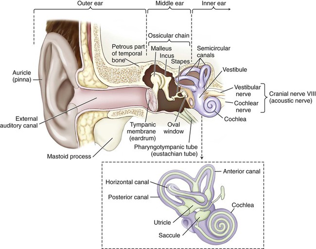
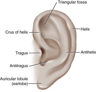
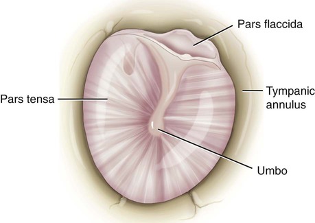
The ear is regionally divided into the outer, middle, and inner ear (Fig. 14-1). Sound travels through air, bone, and fluid across these divisions. Sound waves are initially gathered by the flesh-covered elastic cartilage of the outer ear called the pinna, or auricle (Fig. 14-2). The auricular cartilage is folded into several distinct structures with separate names. The helix is the upper outer rim of the auricle, whereas the antihelix is the inner curve that is parallel to the helix. The antihelix has two “legs,” or crura (sing. crus), that divide to form a shallow depression between them, referred to as the triangular fossa. The tragus is the fleshy tag of tissue with a tuft of hair on its underside that covers the opening of the external auditory canal. The antitragus is the small raised prominence that is opposite to the tragus. It is important to remember that elastic cartilage is covered by a layer of connective tissue called perichondrium. When this is separated from the cartilage by trauma, deformities of the pinna may occur. The lobule, usually referred to as the earlobe, is the only noncartilaginous part of the external ear. This fleshy protuberance is composed of adipose tissue. The tympanic membrane (TM), or eardrum, marks the end of the external ear and the beginning of the middle ear (Fig. 14-3). This concave membrane of the eardrum is attached to an almost complete ring of bone called the tympanic annulus. The membrane is composed of a thick, taut part (the pars tensa) and a thin, flexible part (the pars flaccida). The center of the membrane is pulled inward, forming a shallow depression termed the umbo. Because the membrane is extremely delicate and vulnerable to perforation and infection, the additional terms naming structures of the eardrum are necessary to specify where the injury occurs. Once sound is conducted to the oval window, it is transmitted to a structure called the labyrinth, or the inner ear. A membranous labyrinth is enclosed within a bony labyrinth. Between the two, and surrounding the inner labyrinth, is a fluid called perilymph. Within the membranous labyrinth is a fluid called endolymph. Hair cells within the inner ear fluids act as nerve endings that function as sensory receptors for hearing and equilibrium. Tiny calcium carbonate crystals called otoliths are attached to these hair cells and act as receptors to aid in balance. The outer, bony labyrinth is composed of three parts: the vestibule, the semicircular canals, and the cochlea. The vestibule and semicircular canals provide information about the body’s sense of equilibrium, whereas the cochlea is an organ of hearing. Within the vestibule, two structures called the utricle and the saccule function to determine the body’s static (nonmoving) equilibrium (Fig. 14-1, inset). A specialized patch of epithelium, called the macula, found in both the utricle and the saccule, provides information about the position of the head and a sense of acceleration and deceleration. The semicircular canals detect dynamic equilibrium or a sense of sudden rotation through the function of a structure called the crista ampullaris. Do not confuse oral, meaning pertaining to the mouth, with aural, meaning pertaining to the ear. Anatomy of the Ear Match the combining forms with the correct parts of the ear. More than one answer may be correct. 13. The mucous membrane–lined structure that connects the ear to the throat is the ___________________. 14. The __________________ is a hard, small projection of the temporal bone full of air cells. 15. The ear is regionally divided into the ________________, ________________, and _________________ ears. 16. Another word for outer ear is _________________ or _______________________________________________. 17. The outer, bony labyrinth is composed of three parts: the _________________, the _____________________, and the ______________________________________________________________. 18. The _________________ and the _________________ function within the vestibule to determine the body’s static (nonmoving) equilibrium. Combining Forms for Anatomy and Physiology of the Ear Terms Related to Congenital Disorders of the Ear (QØØ-Q99) Terms Related to Diseases of the External Ear (H6Ø-H62)
Ear and Mastoid Process
 Recognize and use terms related to the anatomy and physiology of the ear.
 Recognize and use terms related to the pathology of the ear.
Anatomy and Physiology
Outer (External) Ear
Inner Ear
 Be Careful!
 Exercise 1:
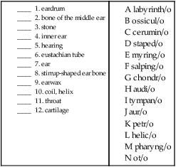
Meaning
Combining Form
antrum
antr/o
auricle, pinna
auricul/o
cartilage
chondr/o, cartilag/o
cerumen, earwax
cerumin/o
cochlea
cochle/o
ear
ot/o
eardrum
tympan/o, myring/o
eustachian tube
salping/o
hearing
acous/o, audi/o, aur/o
helix, coil
helic/o
labyrinth, inner ear
labyrinth/o
mastoid process
mastoid/o
ossicle, small bone
ossicul/o
petrous bone
petros/o
spot, macula
macul/o
stapes
staped/o
stone
petr/o, lith/o
vestibule
vestibul/o
Pathology
Term
Word Origin
Definition
macrotia
macro- large
ot/o ear
-ia condition
A condition of abnormally large auricles.
microtia
micro- small, tiny
ot/o ear
-ia condition
A condition of abnormally small auricles.
Term
Word Origin
Definition
cholesteatoma, external ear
chol/e bile
steat/o fat
-oma mass, tumor
Cystic mass composed of epithelial cells and cholesterol. Can occur also in middle ear (Fig. 14-4).
exostosis of external ear
ex- out
oste/o bone
-osis abnormal condition
Bony growth usually due to chronic irritation.
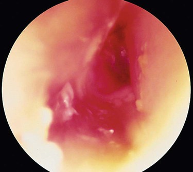
otitis externa
ot/o ear
-itis inflammation
extern/o outer
-a noun ending
Inflammation of the pinna/auricle (Fig. 14-5).
perichondritis, auricular
peri- around, surrounding
chondr/o cartilage
-itis inflammation
Inflammation of the perichondrium of the external ear. May result in a deformity referred to as “cauliflower ear.”
stenosis of external ear canal, acquired
stenosis abnormal condition of narrowing
A narrowing of the auditory canal that develops after birth. ![]()
Stay updated, free articles. Join our Telegram channel

Full access? Get Clinical Tree


Basicmedical Key
Fastest Basicmedical Insight Engine

 Note
Note Be Careful!
 Be Careful!
 You can review the anatomy of the ear by clicking on Body Spectrum, then Senses.
 To practice labeling the parts of the ear, click on Label It.
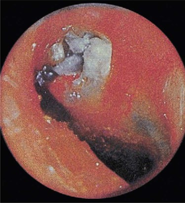

 CM Guideline Alert