Low-power examination shows a dermal-based clear cell tumor with a nonencapsulated lateral border  . (Courtesy A. Lazar, MD, PhD.)
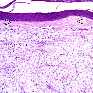
Higher magnification examination shows a dermal-based clear cell tumor with a thin grenz zone
 separating it from the overlying epidermis. (Courtesy A. Lazar, MD, PhD.)
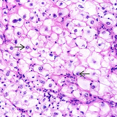
High magnification shows the relatively bland cytologic features of the large clear cells with vesicular nuclei, focally enlarged nucleoli
 , and abundant clear cytoplasm. (Courtesy A. Lazar, MD, PhD.)
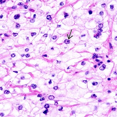
Higher magnification shows relatively bland cytologic features of large clear cells, which show enlarged nuclei with vesicular chromatin and focally prominent nucleoli
 . Cytoplasm is predominantly clear to focally granular-appearing. (Courtesy A. Lazar, MD, PhD.)
CLINICAL ISSUES
Epidemiology


Treatment
• Surgical approaches


Stay updated, free articles. Join our Telegram channel
Full access? Get Clinical Tree
