Digital Fibromatosis (Infantile Digital Fibromatosis)
No nuclear β-catenin labeling but cytoplasmic labeling common
• Inclusions show granular &/or filamentous features by EM
• Cytoplasmic filaments extend onto inclusions
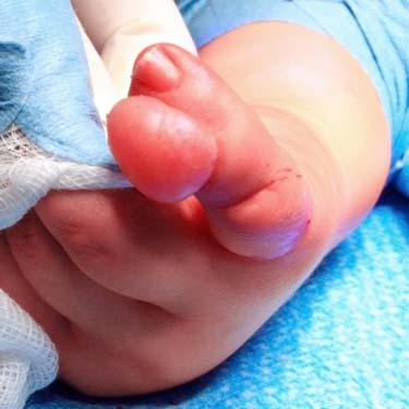
Infantile Digital Fibromatosis/Inclusion Body Fibromatosis Clinical examination of infantile digital fibromatosis shows an exophytic, dome-shaped superficial neoplasm, which presents in infants and small children.
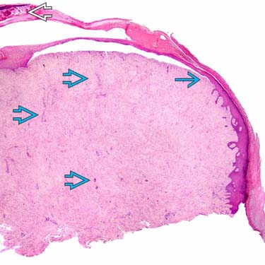
Infantile Digital Fibromatosis, Low Magnification At low magnification, the lesion is plastered against the overlying skin . Because it is pale, tiny capillaries are readily apparent even at low magnification. This example has a surface erosion with exudate .
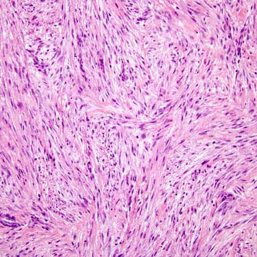
Infantile Digital Fibromatosis, Storiform Architecture Note the storiform pattern of this tumor at intermediate magnification. The process is myofibroblastic, so it is not as brightly eosinophilic as a smooth muscle tumor.
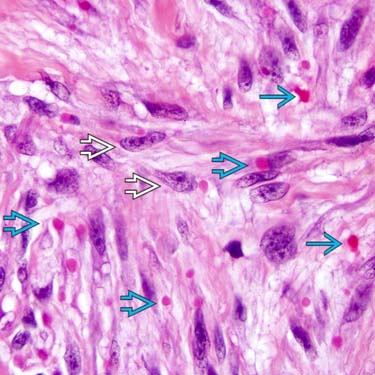
Infantile Digital Fibromatosis, Cytologic Features Note the eosinophilic inclusion bodies , which consist of actin filaments. They are not as eosinophilic as erythrocytes . This image also shows the delicate nuclear chromatin in the myofibroblastic cells of the lesion. Most of the nuclei display delicate nucleoli.
TERMINOLOGY
Synonyms
• Infantile digital fibromatosis
Infantile digital fibroma
• Digital fibrous tumor of childhood
Recurring digital fibrous tumor of childhood
• Inclusion body fibromatosis (term used by World Health Organization)
Definitions
• Benign proliferation of fibroblasts and myofibroblasts, containing scattered eosinophilic spherical inclusions, that arises on digits (both fingers and toes) of young children
CLINICAL ISSUES
Epidemiology
• Incidence
Rare fibroblastic/myofibroblastic neoplasm
• Age
Most cases occur in 1st year of life, occasional cases in older children
Very rare in adult patients
• Sex
M = F
Site
• Dorsal aspects of hands or feet most common
• Rarely synchronous or asynchronous involvement of > 1 digit
• Thumb or great toe is only very rarely affected
• Extradigital soft tissues (i.e., arm, breast) are only extremely rarely affected
Presentation
• Digital enlargement
• Dome-shaped swelling overlying phalanges or interphalangeal joints
• Nontender nodules
• Rarely erosion of bone
Natural History
• May recur locally
• May regress spontaneously
• No progression
• No metastases
Treatment
• Surgical approaches
Local excision with preservation of function
Prognosis
• Excellent overall prognosis
• May recur locally
• May show spontaneous regression
• Main prognostic indicator is adequacy of primary excision
MACROSCOPIC
General Features
• Ill-defined neoplasm
• Dermal-based neoplasm with gray-white, indurated cut surface covered by intact skin
• No areas of hemorrhage
• No areas of necrosis
Size
• Nodules of variable size
Usually measure < 2 cm
MICROSCOPIC
Histologic Features
• Dermal tumor composed of infiltrating fascicles and sheets of spindle cells
• Uniform-appearing spindle-shaped fibroblasts and myofibroblasts
 . Because it is pale, tiny capillaries
  are readily apparent even at low magnification. This example has a surface erosion with exudate
  .
.
 , which consist of actin filaments. They are not as eosinophilic as erythrocytes
  . This image also shows the delicate nuclear chromatin in the myofibroblastic cells
  of the lesion. Most of the nuclei display delicate nucleoli.


