Desmoplastic Fibroblastoma


Desmoplastic Fibroblastoma
Low-magnification examination of a desmoplastic fibroblastoma is shown. The lesion is a well-circumscribed ovoid or fusiform mass. The characteristic hypocellularity is apparent at low power.


image
Desmoplastic Fibroblastoma With Deep Extension
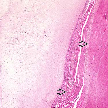
Although most desmoplastic fibroblastomas are subcutaneous, some involve skeletal muscle image, to which this lesion closely abuts.

image
Fibroblastoma With Spindled and Stellate Fibroblasts
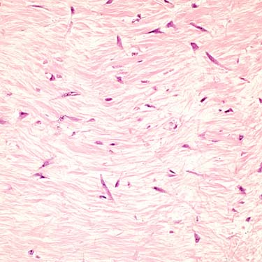
High magnification shows stroma that is densely collagenous and contains spindle and stellate fibroblasts in patternless distributions. The vessels are inconspicuous.

image
Fibroblastoma Myxoid Stroma
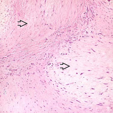
This image of desmoplastic fibroblastoma shows the sparsely cellular nature of the lesion. The stroma can be myxoid image in some cases. Although cells may be plump, no true atypia is seen, and mitotic figures are rare or absent.


TERMINOLOGY


Synonyms


• Collagenous fibroma


Definitions


• Rare benign fibrous soft tissue neoplasm occurring mainly in men and consisting of paucicellular arrays of stellate and spindle fibroblasts


ETIOLOGY/PATHOGENESIS


Developmental Anomaly


• Etiology unknown


CLINICAL ISSUES


Epidemiology


• Age
image All ages, but particularly older adults (5th-6th decades)

• Sex
image M > F


Site


• Most occur in subcutaneous tissue
image Up to 25% involve skeletal muscle

• Most common in upper extremity (shoulder, upper arm, forearm), followed by lower extremity

• Rare in head and neck


Presentation


• Slow growing

• Painless mass


Treatment


• Simple complete excision

Apr 24, 2017 | Posted by in PATHOLOGY & LABORATORY MEDICINE | Comments Off on Desmoplastic Fibroblastoma

Full access? Get Clinical Tree

Get Clinical Tree app for offline access