Psoriasis
Psoriasis is a relatively common disorder affecting 1–2% of the population. Classically it presents with red, raised, scaly patches or plaques, which reflect increased keratinocyte proliferation within the epidermis, associated with an inflammatory cell infiltrate (including polymorph microabscesses). There is also increased vascularity within the upper dermis.
The aetiology of psoriasis remains relatively poorly understood. Although there is clearly a genetic component, with younger patients in particular often reporting a positive family history, patterns of inheritance vary and further studies are ongoing to try to understand which genetic factors are at play. In addition, it remains unclear why some areas of skin are affected, but others remain normal in the face of an underlying genetic predisposition. Triggers that can be associated with the development of/flare-up of psoriasis in susceptible individuals include infections and trauma, and possibly stress, although the latter is contested by some dermatologists.
Clinical Presentation
Several different patterns of psoriasis are recognised, some of which are common, while others are only rarely seen. The key clinical features of each subtype are shown in Table 19.1 and Plates 19.1–19.4.
Table 19.1 Clinical Features and Specific Treatments for Different Types of Psoriasis
| Type | Clinical features | Specific treatment(s) |
| Classical plaque psoriasis (Plate 19.1) | • Single or multiple red plaques, ranging from a few millimetres to several centimetres in diameter | • First choice options include vitamin D analogues, dithranol, topical corticosteroids (± tar or salicylic acid) |
| • Scaly surface – gentle scraping yields a silvery appearance; more vigorous rubbing may result in focal haemorrhage (Auspitz sign) | • UV radiation therapy may be helpful | |
| • Plaques most commonly develop on extensor surfaces – elbows, knees – but may affect any part of the body – typically in a symmetrical manner | • For more severe cases consider PUVA, cytotoxics, retinoids or biological agents | |
| • Most plaques are chronic and stable, although some will evolve/coalesce slowly over time, while others may disappear. New lesions may develop at sites of trauma (Köbner phenomenon) | ||
| • The scalp and nails are also commonly involved (see below) | ||
| Scalp psoriasis (Plate 19.2) | • Commonly coexists with classical plaque psoriasis, but may occur in isolation | • Tar shampoos/gels may be effective (± salicylic acid) |
| • Varies from one or two isolated plaques to a thick scaly sheet covering the whole scalp | • Topical corticosteroids (± salicylic acid) can also be used | |
| Nail psoriasis (Plate 19.3) | • Pitting – typically large/irregular | • Often difficult to treat with topical agents; however, use of systemic agents is seldom justified |
| • Onycholysis – separation of the nail plate from the nail bed; often begins as a small area of red/brown discolouration, but may spread to involve the whole nail | ||
| Guttate psoriasis (Plate 19.4) | • Typically presents suddenly with a ‘shower’ of small round plaques, often on the trunk | • First choice therapy is UV radiation (± tar, emollients) |
| • May develop after an infective episode (especially streptococcal sore throat) | • Topical vitamin D analogues and corticosteroid therapy (often in combination) may be effective | |
| • More likely to be itchy than other forms of psoriasis | ||
| • Lesions may regress rapidly even without treatment | ||
| Flexural psoriasis | • May accompany plaque, scalp or nail psoriasis or occur in isolation | • Difficult to treat: tar-based therapies and topical corticosteroids may help, but long-term use can lead to skin atrophy/striae |
| • Affects groin, axillae, natal cleft, submammary folds | • Low-strength dithranol or vitamin D analogues can be tried but often cause local stinging/irritation | |
| • Maceration of the skin can result in marked redness and loss of the typical scaly appearance | ||
| • Often itchy | ||
| Brittle psoriasis | • Thinner scales | • Requires careful management under expert supervision |
| • May develop in a patient with previously stable plaque psoriasis | • Emollients may be tried, but PUVA or systemic therapy (e.g. methotrexate or retinoids) is often required | |
| • Rapid generalisation/coalescence of lesions leads to erythroderma or acute pustular psoriasis (see below) | ||
| • Episodes may be triggered by use of potent topical or systemic corticosteroid therapy | ||
| Erythrodermic psoriasis | • Occurs when plaques merge to involve most or all of the skin | • Requires urgent dematological review; may become life-threatening if treatment is delayed |
| • May develop rapidly and occasionally arises de novo | • Methotrexate or ciclosporin are effective | |
| • Biological agents are finding increasing use | ||
| Acute pustular psoriasis | • Characterised by widespread erythema with sterile pustules, which may coalesce | • As for erythrodermic psoriasis |
| • Associated with systemic upset, including fever | ||
| • Secondary bacterial infections may be life-threatening if not treated promptly | ||
| Chronic palmo- plantar pustulosis | • Erythematous patches with multiple pustules, which dry to form circumscribed brown areas that eventually peel off | • Difficult to treat; topical measures are usually ineffective |
| • May affect only a small area of one hand or foot or involve the entire surface of both palms and soles | • PUVA or oral retinoid therapy may be tried, but relapses are common |
Psoriatic Arthropathy
Arthropathy occurs in 5–10% of patients with psoriasis and may take one of several forms:
- predominant distal interphalangeal joint involvement
- symmetric polyarthritis (seronegative rheumatoid-like changes)
- asymmetric oligo/pauciarticular arthritis
- spondylitis (± sacro-ileitis) with stiffness in the back/neck; HLA B27 positive
- arthritis mutilans, a severe deforming destructive arthritis.
Nail changes are common in patients with psoriatic arthropathy, but not all cases are associated with skin disease.
Treatment
Topical Agents
- Emollients help to control scaling.
- Salicylic acid reduces hyperkeratotic, scaling lesions; often used in combination with coal tar, dithranol or topical corticosteroids.
- (Coal) tar is effective but unpleasant to use; typically reserved for scalp involvement; sometimes used in combination with ultraviolet (UV) radiation therapy (see below).
- Dithranol is effective for chronic plaque psoriasis; applied directly to lesions (may be covered with a dressing) and left for up to an hour (occasionally longer, but only under supervision); dithranol is irritant (start with 0.1% and gradually increase as required/tolerated) and stains skin, hair and bedding/clothes brown; avoid use on the face or in flexures; may be combined with tar and UV radiation.
- Topical corticosteroids may suppress (although not erradicate) lesions (e.g. scalp, flexures), but should be used with care (risk of precipitating brittle or erythrodermic psoriasis) (Table 19.1).
- Vitamin D analogues (e.g. calcipotriol, tacalcitol) and activated vitamin D (e.g. calcitriol) are useful in mild to moderate psoriasis; serum calcium levels must be monitored in those using larger doses.
- Tazarotene is a retinoid; irritant, especially if applied to normal skin.
Phototherapy
- UVB radiation is effective in the treatment of chronic stable psoriasis and guttate psoriasis, especially when topical agents have proved ineffective; must be used with care due to risk of sunburn; typically given as short courses, e.g. 2–3 times per week until clearance is achieved; combination with coal tar, dithranol or vitamin D analogues increases efficacy.
- Photochemotherapy (psoralens and ultraviolet A phototherapy (PUVA)). Psoralens (given either by mouth or topically) enhance the effect of UV radiation; treatment is typically given twice weekly, and protective glasses are required to avoid ocular damage. Higher cumulative doses exaggerate skin ageing and are associated with an increased risk of keratoses and neoplastic lesions (especially squamous carcinoma).
Systemic Therapy
- Methotrexate. Once weekly dosing, together with folic acid; effective in severe psoriasis refractive to topical therapy; patients should be advised of potential adverse effects including myelosuppression (sore throat, mouth ulceration, easy bruising), hepatotoxicity (nausea/vomiting, abdominal pain, dark urine), respiratory effects (shortness of breath), inhibition of spermatogenesis and teratogenicity (effective contraceptive measures must be in place before treatment is commenced).
- Retinoids (e.g. acitretin) are reserved for severe, resistant psoriasis. Side effects include dryness and cracking of skin and lips, epistaxis, transient hair loss, myalgia, hepatotoxicity, hyperlipidaemia and teratogenicity (effective contraceptive measures must be in place before treatment is commenced).
- Ciclosporin (cyclosporin) is effective even in very severe psoriasis; nephrotoxic and requires close monitoring of renal function, especially in the early stages of treatment; avoid concomitant UVB/PUVA therapy.
- Biological agents. Anti-TNFα therapies (e.g. etanercept, adalimumab, infliximab) may be used in very severe plaque psoriasis that has failed to respond to systemic treatments and to photo(chemo)therapy, or in cases where standard treatments are not tolerated/contraindicated. Ustekinumab (a monoclonal antibody that targets interleukins 12 and 23) has also shown benefit.
- Systemic corticosteroids. Only rarely indicated and must be given under expert supervision.
Table 19.1 shows preferred treatment options for each subtype of psoriasis. Psoriatic arthropathy often responds to simple anti-inflammatory agents. In more severe cases, methotrexate or biological agents can be tried.
Eczema/Dermatitis
Although the term ‘dermatitis’ is sometimes used specifically to describe skin inflammation caused by an exogenous agent, it is in fact synonymous with the term ‘eczema’ (Greek, meaning ‘boiling over’). Eczema is common, affecting approximately 10% of the population at some stage during life.
Clinical Presentation
In acute eczema the skin is erythematous and oedematous, with papules/vesicles and weeping (i.e. ‘boiling over’). In chronic eczema, oedema is absent and the epidermis becomes thickened/hyperplastic, with exaggeration of the skin markings (so-called lichenification) (Plate 19.6). Itching may be the dominant symptom. Secondary infection is common: bacterial (commonly staphylococcal) or viral (herpes simplex). Healed lesions do not scar, but may pigment.
Secondary spread to involve other areas is a well-recognised phenomenon of acute eczema and may even involve areas that have not been directly exposed to a particular allergen in cases of contact dermatitis. Rarely, the whole of the body may be affected by a generalised exfoliative dermatitis.
Several different patterns of eczema are recognised; the key clinical features of each subtype are shown in Table 19.2 and Plates 19.5–19.7.
Table 19.2 Causes, Clinical Features and Specific Treatments for Different Types of Eczema (Dermatitis)



Treatment
Wherever possible, the local irritant or sensitiser should be removed. Many commercial soaps contain such irritants and washing with water ± soap substitutes is advised. If the lesion is weeping, local soaks, e.g. potassium permanganate (antiseptic and astringent) may aid healing; if dry, emollients should be applied liberally and regularly to the affected areas – bath/shower oils may further help. Continued emollient use is required even when the acute episode has subsided. Sedative antihistamines by mouth may relieve pruritus and allow sleep. Bandages (including those containing zinc and ichthammol) may be applied over topical corticosteroids (see below) or emollients to treat eczema of the limbs. Systemic antibiotics are required for secondary bacterial infections and antiviral therapy for cases complicated by herpes simplex infection. Antifungal therapy may be useful in cases of seborrhoeic dermatitis where the yeast Malassezia is implicated.
Topical corticosteroids remain the mainstay of treatment for most patients with eczema. The potency of the corticosteroid should be appropriate for the site and severity of the condition, e.g. weaker preparations are preferred for the face and on flexures, while more potent preparations may be required on the limbs/trunk, especially if there is associated lichenification.
In more severe/refractory cases immunomodulatory agents can be tried, including topical tacrolimus or pimecrolimus and oral ciclosporin. The oral retinoid alitretinoin is also licensed for the treament of severe chronic hand eczema refractory to potent topical corticosteroids (cautions and adverse effects are similar to those of other retinoid preparations; pregnancy must be excluded and effective contraception practised).
Table 19.2 shows preferred treatment options for each subtype of eczema.
Acne Vulgaris
Acne vulgaris is a disease chiefly of puberty (but onset can occur up to 40 years of age, and even at later ages if there is an underlying endocrine disorder, e.g. Cushing syndrome) in which androgens (typically in normal amounts) promote increased sebaceous gland activity, leading to greasy skin. Hyperkeratosis results in plugging of hair follicles, and subsequent secondary infection with the obligate anaerobe Propionibacterium acnes leads to release of chemicals into the surrounding dermis, which provoke an intense inflammatory response.
Clinical Presentation
Several different lesions may be seen:
- Comedones – closed (‘whitehead’) or open (‘blackhead’)
- Papules and pustules – spots or pustules on a red base; may recur repeatedly in the same locations
- Nodules and cysts – seen in more severe cases, as inflammation extends deeper; if these are numerous they are referred to as ‘acne conglobata’; very rarely nodulocystic acne is associated with fever, malaise and systemic upset (‘acne fulminans’)
- Scars – these may be pitting and disfiguring, and in extreme cases associated with keloid formation.
Investigation
In the majority of patients investigation is not required, but disorders associated with hyperandrogenism should be considered in female subjects with later onset disease or in the presence of virilising features (e.g. Cushing syndrome, androgen-secreting tumours).
Treatment
Options include:
- over-the-counter preparations – effective in milder cases and work mainly as astringents/keratolytics, drying the skin and unblocking hair follicles
- topical benzoyl peroxide – astringent, keratolytic and delivers oxygen locally, reducing bacterial proliferation without inducing resistance; may cause local skin irritation
- topical antibiotics – e.g. tetracyclines, erythromycin, clindamycin; effective in the early stages of treatment, but resistance often develops
- topical retinoids – e.g. isotretinoin, adapalene; reduce sebum production and attenuate inflammation
- oral antibiotics – e.g. tetracyclines, erythromycin; both antibacterial and anti-inflammatory
- oral isotretinoin (Roaccutane®) – shrinks sebaceous glands, dramatically reducing sebum production; side effects are common, including facial erythema, dry skin and lips, nose bleeds, myalgia, hyperlipidaemia and abnormal liver function tests. It is teratogenic: pregnancy must be excluded and reliable contraception established before commencing treatment.
Patients should be warned that most treatments take several months of continued use before benefits become apparent.
It is important to tackle underlying disorders when present (e.g. promoting weight loss in overweight/obese females with polycystic ovarian syndrome; treating Cushing syndrome).
The role of UV light therapy in treating active acne and healing scar tissue is currently under investigation.
Rosacea
This is a disorder, more common in women, beginning usually after 30 years of age, with erythema, papules, pustules and telangiectasia over the cheeks, nose, chin and forehead. It may be mistaken for acne, but there are no comedones. Flushing is common, especially in warm environments or in response to alcohol. In men, sebaceous hyperplasia on the nose leads to the condition ‘rhinophyma’.
Oral tetracyclines are preferred for treatment but as with acne take several months to produce maximum benefits. Topical metronidazole may also be added. Oral isotretinoin can be effective in more resistant/severe cases. Plastic surgery may be required for rhinophyma. Precipitating factors should be avoided (e.g. hot drinks, warm environments, alcohol, sunlight, topical corticosteroids).
Hidradenitis Suppurativa
A rare disorder characterised by relapsing suppurative infection in the axillae and groins – sites at which apocrine glands open into pilosebaceous follicles. It is more commonly seen in females, and flare-ups may correlate with changes in hormonal levels, e.g. in the menstrual cycle. Obesity exacerbates, but does not cause, the condition. Recurrent painful abscesses and sinus tracks develop, which discharge unpleasant material. Although oral antibiotics and oral isotretinoin can be tried, success is usually limited and surgical intervention is often required to lay open the sinus tracks and excise chronically infected areas.
Bacterial, Viral, Fungal and Parasitic Skin Infections
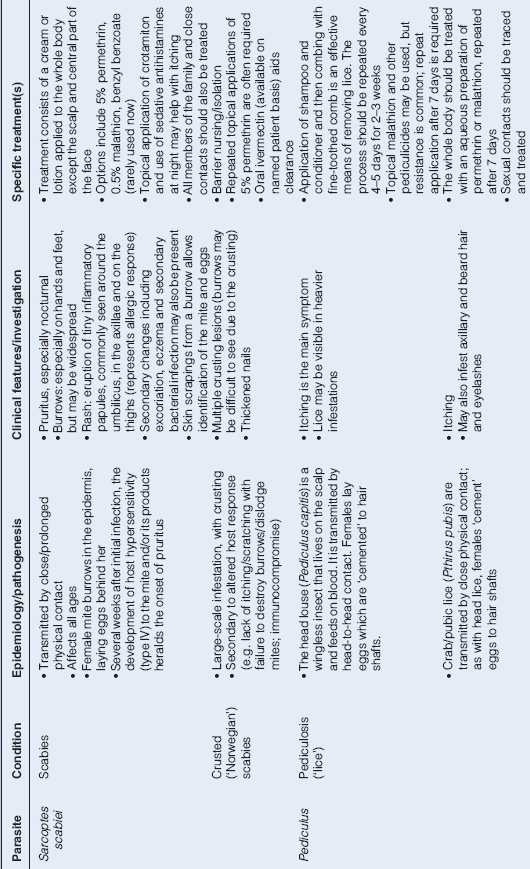
A number of different organisms are capable of causing primary cutaneous infections/infestations. The more commonly encountered pathogens and their associated clinical sequelae are shown in Tables 19.3–19.6 and Plate 19.8.
Table 19.3 Bacterial Infections Involving the Skin.


Table 19.4 Viral Infections Involving the Skin.


Table 19.5 Fungal Infections Involving the Skin.


Table 19.6 Parasitic Infections Involving the Skin.
Cutaneous Drug Reactions
Cutaneous drug reactions are relatively common and may arise through one of several different mechanisms:
- intolerance
- hypersensitivity (types I, II, III and IV)
- interactions with other drugs
- interactions with other environmental or host factors (e.g. sunlight exposure)
- pharmacokinetic disturbances
Table 19.7 show the different types of eruption that may be encountered. Drugs that are particularly prone to causing cutaneous reactions include antibiotics (e.g. sulphonamides, penicillins – although not all patients who state they are ‘penicillin allergic’ are truly allergic!), NSAIDs and hypnotics/tranquillisers.
Table 19.7 Patterns of Cutaneous Drug Reactions
| • Exanthematous eruptions |
| • Urticaria/anaphylaxis |
| • Eczema |
| • Exfoliative dermatitis |
| • Fixed drug eruptions |
| • Vasculitis |
| • Erythema multiforme |
| • Erythema nodosum |
| • Pigmentary changes |
| • Bullous/blistering reactions |
| • Photosensitivity |
| • Acneiform eruptions |
| • Hair loss or hair growth |
| • Lupus erythematosus-like syndrome |
| • Lichen planus-like eruptions |
| • Purpura |
| • Exacerbation of pre-existing skin disorder (e.g. acne, psoriasis, systemic lupus erythematosus, porphyria) |
Key features of the major types of drug reaction are described below.
Clinical Features/Causative Agents
Exanthematous Eruptions
- variable speed of onset – typically starts within the first few days of treatment, but may develop more quickly (e.g. within hours of exposure) or appearance may be delayed (e.g. several weeks, making the link more difficult to establish)
- usually widespread, symmetrical, erythematous maculopapular rash, often resembling a viral exanthem (Plate 19.9)
- itchy
- examples: penicillins, sulphonamides, NSAIDs
Urticaria and Anaphylaxis
- secondary either to direct effect on mast cells or type I or type III hypersensitivity reaction
- eruption usually occurs 3–7 days after therapy is started
- rarely, rapid onset anaphylactic reactions (with fever, wheezing, arthralgia and hypotension) occur and are potentially fatal if not rapidly recognised and treated
- examples: penicillins, cephalosporins, aspirin, opioids, certain vaccines
Eczema
- type IV hypersensitivity reaction
- typically in response to a topical agent
- examples: lanolin, preservatives, topical antibiotics (especially aminoglycosides), topical antihistamines, topical local anaesthetics (although not lignocaine), topical corticosteroids
Exfoliative Dermatitis (Erythroderma)
- widespread reddening/inflammation of the skin ± scaling
- in more severe cases may be associated with loss of temperature regulation, dehydration, superadded infection, hyperdynamic circulation (± cardiac failure in the elderly)
- examples: phenytoin, sulphonylureas, sulphonamides, allopurinol, gold, barbiturates
Fixed Drug Eruptions
- typically manifests as a circular/oval patch of erythema with central purple discolouration ± bullous change
- single or multiple sites
- often initially misdiagnosed as ringworm infection
- usually affects the same area(s) of skin each time the patient is exposed to the offending drug
- postinflammatory pigmentation
- examples: phenolphthalein-containing laxatives, tetracyclines, sulphonamides, dapsone
Vasculitis (p. 284)
- typically a cutaneous small vessel vasculitis (leukocytoclastic vasculitis)
- numerous palpable, purpuric lesions (often on the lower limbs); occasionally developing into haemorrhagic vesicles/bullae
- may be associated with systemic involvement (e.g. renal)
- examples: thiazide diuretics, cimetidine, sulphonamides
Erythema Multiforme (Plate 19.10)
- target lesions
- examples: sulphonamides, cotrimoxazole, rifampicin, barbiturates
Erythema Nodosum
- tender, red, raised lesions (Plate 19.11)
- typically on the shins, but may also affect upper limbs
- examples: sulphonamides, salicylates
Pigmentary Changes
- various drugs can be associated with different pigmentary skin changes
- examples: blue-black (chloroquine, amiodarone (in sun-exposed areas)); brown (oestrogens = chloasma)
Bullous/Blistering Reactions
- may be seen in the context of a fixed drug eruption or with drug-induced pemphigus, pemphigoid or porphyria cutanea tarda
Photosensitivity
- direct phototoxic effect or exacerbation of a pre-existing condition
- in phototoxic reactions, the dose of the drug and the degree of ultraviolet radiation exposure determine the extent of the reaction; characterised by erythema, swelling ± eczematous changes
- examples: tetracyclines, sulphonamides, phenothiazines, thiazide diuretics
Acneiform Eruptions
- papules/pustules
- examples: corticosteroids, androgenic drugs, lithium
Lupus-Erythematosus-Like Syndrome
- rare
- examples: hydralazine, isoniazid, minocycline
Lichen Planus-Like Eruptions
- rare
- may be indistinguishable from idiopathic lichen planus, although eczematous/scaly changes are more common
- examples: antimalarials, sulphonylureas, gold, thiazide diurectics (in sun-exposed areas)
Purpura
- may be a feature of any severe drug reaction and typically results from capillary damage and/or thrombocytopenia
Management
- Wherever possible discontinue suspected offending drug.
- Minimise other potential provoking factors, e.g. UV radiation exposure.
- Oral antihistamines for urticaria.
- Mild topical steroids may help itching.
- Adrenaline (epinephrine) may be life-saving in acute hypersensitivity reactions including shock and angioedema (p. 115), and systemic steroids may be required in severe but less acute cases.
Skin Manifestations of Systemic Disease
Skin involvement in systemic disease is not uncommon and can be the presenting feature, e.g. erythema nodosum in sarcoidosis (p. 120). In some instances, several different underlying disorders can give rise to the same skin condition (Table 19.8), while other cutaneous manifestations are more specific. Some disorders (e.g. diabetes mellitus) are associated with a wide array of cutaneous features.
Table 19.8 Skin Conditions Associated with a Variety of Systemic Diseases
| Cutaneous manifestation | Associated systemic disease(s) | Clinical features/treatment |
| Erythema nodosum (Plate 19.11) | • Sarcoidosis | • Tender, red, raised areas, typically on the shins but occasionally on the forearms |
| • Streptococcal infection | • With time the lesions pass through the colour changes of a bruise before resolving | |
| • Tuberculosis | • Simple analgesia usually suffices in the acute phase | |
| • Inflammatory bowel disease | ||
| • Systemic fungal infections | ||
| Erythema multiforme (Plate 19.10) | • Herpes simplex infection | • Target lesions, typically over extensor surfaces of arms and legs, but may spread to involve other areas of the body; dusky purplish centre which may blister |
| • Mycoplasma infection | • Self-limiting in most cases | |
| • Less commonly: connective tissue disorders; malignancy | • Occasionally associated with major systemic upset (Stevens–Johnson syndrome), with lesions in the mouth, conjunctiva and anogenital regions; treatment is supportive (the role of systemic corticosteroids remains controversial) | |
| Pyoderma gangrenosum (Plate 19.14) | • Inflammatory bowel disease | • Necrotic ulceration with characteristic bluish/purplish undermined edge |
| • Rheumatoid arthritis | • Single or multiple lesions, usually on the lower limb | |
| • Seronegative arthritis | • Painful | |
| • Paraproteinaemia | • Treatment of the underlying condition, with judicious use of systemic corticosteroids; azathioprine and ciclosporin may also be effective |
Infections
- haemolytic streptococcal infection – erythema nodosum (Table 19.8, Plate 19.11), erythema multiforme (Table 19.8, Plate 19.10), erythema marginatum
- acquired immune deficiency syndrome (AIDS) – oral/oesophageal candidiasis, ‘hairy leukoplakia’ (Epstein–Barr virus), seborrhoeic dermatitis, perianal warts, recurrent/severe herpes simplex, Kaposi’s sarcoma, lipodystrophy (in those on highly active antiretroviral therapy)
- Lyme disease (due to infection with the spirochaete Borrelia burgdorferi; transmitted by Ixodid tick bite) – erythema chronicum migrans
Diabetes Mellitus
Skin manifestations include:
- neuropathic and neuroischaemic ulcers
- xanthomata – signifying hyperlipidaemia
- lipohypertrophy (rarely lipoatrophy)
- necrobiosis lipoidica – typically occurs on the shins; intially erythematous, but becomes yellowish brown and atrophic with visible vessels beneath the skin; occasionally ulcerates (Plate 19.12)
- diabetic dermopathy – small brown scar-like lesions, often on the shins
- acanthosis nigricans – indicating the presence of severe insulin resistance (Plate 19.13)
- cheiroarthropathy – thickening of the skin of the hands
- mucosal candidiasis (e.g. balanitis, vulvovaginitis)
- granuloma annulare.
Endocrine Disorders
- thyrotoxicosis/hyperthyroidism – palmar erythema, alopecia; Graves’ disease is specifically associated with acropachy (digital clubbing), onycholysis, pretibial myxoedema
- hypothyroidism – pallor, malar flush (which together may lead to the classical ‘strawberries and cream’ appearance), thinning of scalp hair, loss of outer part of eyebrows, thickened/dry skin (myxoedema)
- Cushing syndrome – thinning of skin, spontaneous/easy bruising, acne, hirsutism, violaceous striae
- Addison’s disease – hyperpigmentation (especially palmar creases, buccal mucosa, scars); may be associated with vitiligo
Hyperlipidaemia
Both primary and secondary hyperlipidaemia may be associated with lipid deposits in the skin (xanthomata), which are yellow/orange in colour and may occur as:
- xanthelasma(ta) – eyelids
- tendon xanthomata – extensor tendons of the hands, Achilles tendons
- palmar xanthomata – creases of the palms
- tuberous xanthomata – over bony prominences
- eruptive xanthomata – crops of papules (indicative of hypertriglyceridaemia).
Rheumatological Disorders
- gout – tophaceous deposits
- rheumatoid arthritis – palmar erythema, rheumatoid nodules, vasculitic lesions, pyoderma gangrenosum
- systemic lupus erythematosus – facial erythema (‘butterfly rash’), photosensitivity, alopecia, Raynaud’s phenomenon
- discoid lupus erythematosus – principally affects light-exposed areas, with scaling and erythematous plaques; healed areas show scarring and hypopigmentation
- dermatomyositis – purple heliotrope discolouration, classically around the eyes, but may involve other areas, especially sun-exposed; periorbital oedema; vasculitic lesions in the childhood variant
- systemic sclerosis – tight, shiny appearance of the skin over the face, with beaked nose and restriction of mouth-opening; facial telangiectasia; sclerodactyly, digital infarcts, calcinosis, Raynaud’s phenomenon
- Reiter syndrome – keratoderma blennorrhagicum, buccal mucosal ulceration, circinate balanitis
Sarcoidosis
Skin manifestations include:
- erythema nodosum (Table 19.8, Plate 19.11)
- lupus pernio – purplish discolouration of the skin of the nose and ears
- papules, nodules, plaques, sarcoid granulomas.
Malignancy
Skin manifestations of malignancy include:
- cutaneous deposits, e.g. breast, bronchus, renal, ovarian (including ‘Sister Joseph’s nodule’, an umbilical metastatic nodule)
- generalised pruritus (associated with a wide array of systemic malignancies, especially lymphoma)
- acanthosis nigricans (Plate 19.13) (gastrointestinal adenocarcinoma)
- dermatomyositis (bronchus, breast, stomach, ovary)
- thrombophlebitis migrans (pancreatic carcinoma)
- flushing (carcinoid syndrome)
- necrolytic migratory erythema (glucagonoma)
- pyoderma gangrenosum (Table 19.8, Plate 19.14) (myeloma)
- acquired ichthyosis (lymphoma).
Miscellaneous Disorders
- liver disease – pruritus, palmar erythema, spider naevi, xanthelasma(ta)
- inflammatory bowel disease – erythema nodosum, pyoderma gangrenosum (Table 19.8, Plate 19.14), buccal mucosal and perianal ulceration
- amyloidosis – yellow waxy periorbital and perianal plaques
- scurvy (due to vitamin C deficiency) – perifollicular purpura, easy bruising, bleeding gums, poor wound healing
- pellagra (due to nicotinic acid deficiency) – triad of dermatitis (in sun-exposed areas, e.g. ‘Casal’s necklace’), diarrhoea and dementia
- neurofibromatosis – café-au-lait spots, axillary freckling, neurofibromas
- porphyria – photosensitive rash/blistering in certain types of porphyria (see p. 254)
- Ehlers–Danlos syndrome – skin hyperextensibility and fragility
- tuberous sclerosis complex (Epiloia) – hamartomas, angiofibromas, shagreen patch, periungal fibromas, hypopigmented (ash leaf) macules
- Peutz–Jeghers syndrome – pigmented macules (lentigines) in the mouth, on the lips, hands and feet
- hereditary haemorrhagic telangiectasia – facial telangiectasia
- pseudoexanthoma elasticum – ‘plucked chicken’ skin appearance
Bullous Disorders
Blisters and bullae can be caused by a wide variety of disorders including physical injury (e.g. friction, extremes of temperature, chemicals, insect bites), infection (e.g. impetigo, varicella zoster), drugs (e.g. sulphonamides, barbiturates), systemic disease (e.g. porphyria) and primary skin conditions. The latter may be congenital (e.g. epidermolysis bullosa) or acquired (e.g. pemphigus, pemphigoid, dermatitis herpetiformis).
Epidermolysis Bullosa
A rare disorder, which presents in the newborn with fragile skin that blisters on minimal contact; may be fatal.
Pemphigus
In pemphigus, splits occur within the epidermis above the basal layer, with degeneration of epidermal cells (acantholysis). Pemphigus vulgaris is the most commonly encountered variant.
Clinical Presentation
Pemphigus is a relatively rare disorder of middle age, some of the characteristics of which are explained by the very superficial site of the lesions: clinically, it presents with widespread erosions and relatively few bullae (because they rupture so easily, leaving flaccid blisters), which are located over the limbs and trunk (Plate 19.15). Most patients have lesions in the mouth and these may be the only visible lesions in the early stages. The surrounding skin is normal. The superficial skin layer at the edge of a blister can be moved over the deeper layers (Nikolsky’s sign) and tends to disintegrate. Lesions appear at sites of pressure and trauma and are painful. Secondary bacterial infection may complicate the primary condition.
Investigation
- skin biopsy – for histopathology (to confirm superficial nature of the blister/bulla) and direct immunofluorescence on perilesional tissue (which shows staining around epidermal cells with antibodies directed against immunoglobulin G (IgG) and C3)
- serum for detection of anti-epithelial antibody
Management
Aggressive management is required including:
- High dose systemic corticosteroids (e.g. prednisolone initially 60–120 mg/day), with gradual dose tapering as blistering settles. Steroid-sparing agents (e.g. azathioprine, cyclophosphamide, methotrexate) are often susbstituted after the acute phase has subsided.
- Secondary bacterial infection is common and should be treated promptly.
- Significant fluid and protein loss may occur from weeping skin, and supportive treatment (including enteral/parenteral feeding in cases of severe oral involvement) may be required.
Pemphigoid
In contrast to pemphigus, blisters are subepidermal.
Clinical Presentation
Bullous pemphigoid affects those > 60 years of age. Clinically, it often presents with prodromal itch ± areas of erythema, which may predate the appearance of bullae by several weeks. Numerous tense, subepidermal bullae then form, ranging in size from a few millimetres to several centimetres (Plate 19.16). They are less likely to rupture than in pemphigus, but this can be provoked by trauma. Nikolsky’s sign is negative. Scarring is rare and only a small number of cases develop mucosal ulceration.
Cicatricial pemphigoid is a distinct variant in which scarring occurs and can be pronounced.
Investigation
- Skin biopsy – for histopathology (to confirm subepidermal blister/bulla). Immunofluorescence shows linear IgG and C3 at the basement membrane).
- Circulating IgG against antigen in the basement membrane is detectable in the serum of approximately two-thirds of patients with bullous pemphigoid.
Management
- Moderate dose systemic corticosteroids (e.g. prednisolone initially 40–60 mg/day) are required, with dose tapering as blistering settles, which usually occurs quite rapidly in bullous pemphigoid.
- Long-term low dose maintenance therapy is often required; azathioprine may be susbstituted after the acute phase has subsided.
Dermatitis Herpetiformis
A rare disorder associated with subepidermal blisters. It is classically seen in the context of coeliac disease.
Clinical Presentation
Dermatitis herpetiformis is characterised by itchy erythematous papules and vesicles, which are common on the elbows and other extensor surfaces. Blisters/bullae may be burst by scratching, with marked excoriation.
Investigation
- skin biopsy – for histopathology (to confirm subepidermal blister/bulla and ‘microabscesses’ at the edge of vesicles) and direct immunofluorescence (which shows granular deposits of IgA in dermal papillae)
Management
- gluten-free diet
- dapsone
Benign and Malignant Skin Tumours
Skin tumours are common. Most are benign, but it is important to identify malignant or potentially malignant lesions. They may arise within the epidermis or the dermis. Clinical features and treatments for the more commonly encounted/important skin tumours are shown in Table 19.9 and Plates 19.17–19.21.
Table 19.9 Skin Tumours
| Tumour | Epidemiology/clinical features | Treatment |
| Benign | ||
| Seborrhoeic keratoses (basal cell papillomas; seborrhoeic warts) | • Common, especially in the elderly | • If required, treatment options range from cryotherapy for smaller lesions to curettage and surgical excision for larger ones |
| • Solitary or multiple | ||
| • Typically occurring on the head, neck, trunk, hands | ||
| • Raised, flat-topped lesions, ranging in colour from light brown to deeply pigmented | ||
| • May be associated with pruritus | ||
| Keratoacanthoma | • Typically seen in elderly people | • Surgical excision and histological examination should be considered for (1) all lesions that cannot be reliably differentiated clinically from a squamous cell carcinoma and (2) persistent lesions |
| • Round with raised edges and characteristic central keratin plug ± reddened/inflamed base | ||
| • Develops over a relatively short time period (2–3 months) and ultimately regresses spontaneously | ||
| Dermatofibroma | • Commonly seen as single or multiple lesions on the lower limbs, especially in females | Usually none required; surgical excision can be considered but may leave scarring |
| • May arise at sites of minor trauma or insect bites | ||
| • Slightly raised, skin-coloured or pigmented | ||
| Pyogenic granuloma (benign proliferation of blood vessels/fibroblasts) | • Polypoidal lesion, which may bleed profusely following minor trauma | • Curettage or surgical excision with histological examination |
| Dysplastic/malignant | ||
| Actinic (solar) keratoses (Plate 19.17) | • Areas of dysplastic squamous epithelium, which typically develop in UV radiation exposed areas (e.g. scalp, face, hands, lower legs) | • Topical application of 5-fluorouracil, imiquimod or a non-steroidal anti-inflammatory cream is generally effective |
| • Dry, rough, scaly lesions, with erythematous background | • Cryotherapy (if lesions limited in size and number) | |
| • May undergo spontaneous involution | ||
| Basal cell carcinoma (BCC; ‘rodent ulcer’) (Plate 19.18) | • Commonest skin cancer | • For superficial tumours, curettage, topical imiquimod, cryotherapy or photo(dynamic) therapy may suffice |
| • UV radiation exposure is an important trigger, and lesions are most commonly seen in sun-exposed areas, e.g. on the face | • Surgical excision and/or radiotherapy are required for deeper lesions | |
| • Often begins as a nodule, which develops a central depression as the lesion extends outwards, leaving a ‘rolled’ edge appearance | ||
| • Contact bleeding from overlying telangiectasia and central ulceration may be seen | ||
| • Locally invasive, but rarely metastasise | ||
| Squamous cell carcinoma in situ (Bowen’s disease) | • Confined to the epidermis, and typically arises in regions subjected to long-term UV radiation exposure (e.g. lower legs), but may also occur in non-exposed areas | • Curettage, cryotherapy, photo(dynamic) therapy or surgical excision may be required |
| • Often presents as a single red, scaly patch, which may be mistaken for psoriasis | ||
| Squamous cell carcinoma (SCC) (Plate 19.19) | • Aetiological factors include UV radiation exposure, smoking in perioral tumours, human papilloma virus infection in genital lesions and immunosuppression (e.g. in transplant recipients) | • Surgical excision and/or radiotherapy |
| • Locally invasive, with greater propensity to metastasise than BCCs | ||
| • Varied appearance: ulcer, keratotic nodule, rapidly expanding polypoidal mass | ||
| Lentigo maligna (‘Hutchinson’s malignant freckle’) (Plate 19.20) | • Patch of malignant melanocytes that have not yet become invasive | • Biopsy to confirm diagnosis followed by surgical excision |
| • Typically develops in UV radiation-damaged skin | • Occasionally surveillance (e.g. in a very elderly patient) may be reasonable | |
| • Flat, brown with variable pigmentation | • Cryotherapy and topical therapies are associated with higher rates of local recurrence | |
| Malignant melanoma (Plate 19.21) | • Worldwide incidence has risen in recent years, especially in younger age groups | • ‘Prevention is better than cure’, e.g. avoidance of strong sunlight, use of ‘sun-blocks’ |
| • UV radiation damage to skin (e.g. repeated sunburn) significantly increases risk | • Patients should be advised to seek early medical advice about suspicious lesions, especially those that develop or enlarge in adult life, if there is an irregular margin or irregular pigmentation, itching or crusting/bleeding | |
| • May arise de novo or in a long-standing mole | • Prognosis is related to the ‘Breslow thickness’ (i.e. depth of tumour at first surgical excision): in essence the thinner the melanoma the better the prognosis (< 1 mm, 95% 5-year survival; > 3.5 mm, < 45% 5-year survival) | |
| • Metastasise to loco-regional and then distant lymph nodes | • Surgical excision remains the mainstay of treatment | |
| • Different patterns are recognised: | • Standard chemotherapy regimens show limited efficacy; radiotherapy may be used for local/distant spread, but generally does not improve prognosis | |
| – lentigo maligna melanoma: malignant nodule developing within a patch of lentigo maligna | • Immunotherapy and agents targeting specific cellular components (e.g. mutated BRAF protein) are under development | |
| – superficial spreading melanoma: irregularly pigmented patch with irregular margins; may itch or bleed | ||
| – nodular melanoma: more rapidly growing; occasionally lacks pigment (‘amelanotic melanoma’) | ||
| – acral melanoma: pigmented patch on the sole or palm or subungal (must be distinguished from a haematoma!) | ||
| Kaposi’s sarcoma | • Multicentric malignant vascular tumour, originally limited to those of Mediterranean and Jewish descent, but now recognised as an AIDS-defining condition | • In the context of AIDS, lesions often resolve in response to HAART |
| • Purple to brown/black plaques/nodules | ||
| HAART, highly active antiretroviral therapy. | ||
Miscellaneous Skin Conditions
Skin Pigmentation
Abnormalities of skin pigmentation are seen in a variety of settings and may be localised to small areas or more generalised. Table 19.10 lists some of the more common causes of hypo- and hyperpigmentation.
Table 19.10 Causes of Hypo- and Hyperpigmentation
| Hypopigmentation |
| • Vitiligo |
| • Pityriasis versicolor |
| • Postinflammatory |
| • Drug/chemical-induced |
| • Leprosy |
| • Lichen sclerosus |
| • Congenital: albinism, phenylketonuria |
| Hyperpigmentation |
| • Addison’s disease |
| • Renal or liver failure |
| • Haemochromatosis |
| • Postinflammatory |
| • Drug/chemical-induced |
| • Chloasma |
| • Acanthosis nigricans |
| • Congenital: neurofibromatosis, Peutz–Jeghers syndrome |
Urticaria
Urticaria describes a group of disorders that are characterised by weals, which typically appear and then disappear spontaneously in a matter of hours. Mast cell degranulation leads to vasodilatation with consequent dermal oedema. Often itching is the first symptom, followed shortly afterwards by the development of pink weals over a variable-sized area, e.g. localised in response to a nettle sting or more generalised as part of an allergic reaction (e.g. food or drug allergies). When part of a more systemic anaphylactic reaction, urticaria may be accompanied by angioedema, with swelling/oedema of the subcutaneous tissues, especially around the eyes, mouth and upper airway. A chronic relapsing form, in which attacks last for weeks, months or even years, is believed to be of autoimmune origin.
If possible, triggers should be identified and avoided. Aspirin is best avoided. Most types of urticaria respond to antihistamines (both H1 and H2 antagonists may be helpful). Angioedema/anaphylaxis are medical emergencies (p. 115).
Lichen Planus
Lichen planus is uncommon, usually presenting in middle age with an irritating rash affecting the flexures of the wrist and forearms, lower back, mouth and genitalia. The rash consists of discrete, purple, shiny, polygonal papules with fine white lines (‘Wickham’s striae’), often occurring in scratch marks and other sites of injury (Köbner phenomenon). The papules may be widespread or confined to one or two sites. Lesions may arise on the buccal mucosa with a white, lacy network, or in the nails without other lesions on the skin. The disorder usually resolves within 6 months but can recur. The cause is unknown, but several drugs can produce an identical eruption, e.g. gold, antimalarials and antituberculous drugs. The epidermis is infiltrated with T cells.
Topical corticosteroids are usually sufficient to suppress symptoms until resolution has occurred. Systemic corticosteroids or ciclosporin may be required for extensive/severe disease.
Pityriasis Rosea
A self-limiting disorder, of unknown aetiology, commonest in children and young adults. Following a mild prodromal illness one or more ‘herald patches’ (red, oval, scaly) appear either on the trunk or arm. Several days later, multiple pink/red oval patches erupt over the trunk, upper arms and thighs, with the long axis of the oval appearing to follow individual dermatomes/spinal roots. It resolves spontaneously over 6–8 weeks.
Disorders of the Hair and Nails
Hair Abnormalities
These generally fall into one or more of three categories:
- Changes in colour or texture (e.g. brittleness, coarseness).
- Thinning or loss of hair – may be congenital or, more commonly, acquired, e.g. telogen effluvium (in which large numbers of hairs suddenly stop growing and enter the ‘telogen’ phase simultaneously; often triggered by stress/intercurrent illness), androgenetic alopecia, alopecia areata (autoimmune disorder, with patchy loss of scalp hair (close inspection at the edge of the patch reveals ‘exclamation mark hairs’); tends to run a relapsing/remitting course; occasionally involves the whole scalp (alopecia totalis) or whole body (alopecia universalis)) and trichotillomania (compulsive plucking of the hair). It may also be seen in the context of skin conditions (e.g. psoriasis, seborrhoeic dermatitis, tinea capitis) and systemic disorders (e.g. hypothyroidism, hypopituitarism) and following drug therapy (e.g. cytotoxic agents)
- Excessive hair growth and development of hair in abnormal sites – hirsutism describes male-pattern hair growth in a female and is most commonly ‘idiopathic’ or associated with the polycystic ovarian syndrome (p. 227), but occasionally can be a sign of a virilising tumour; hypertrichosis is excessive hair growth in a non-sexual distribution and may occur in both sexes (e.g. in response to drugs such as minoxidil and ciclosporin).
Nail Abnormalities
Nail disorders may occur in isolation or may be a sign of more generalised skin disease (e.g. psoriasis, fungal infections) or of an underlying systemic disorder (e.g. koilonychia in iron deficiency anaemia). They include:
- Beau’s lines (horizontal lines following major illness)
- brittleness
- clubbing
- koilonychia (‘spoon-shaped’)
- onychogryphosis (gross thickening)
- onycholysis (lifting of the nail plate off the nail bed, e.g. in psoriasis, Graves’ disease)
- paronychia
- pitting (e.g. in psoriasis)
- Pterygium (e.g. in lichen planus).
Stay updated, free articles. Join our Telegram channel

Full access? Get Clinical Tree


