Patient Story
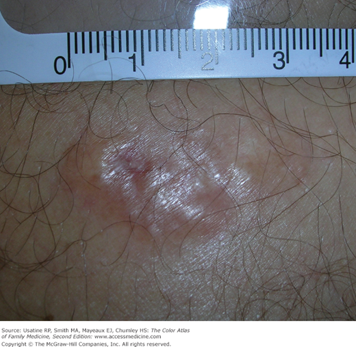
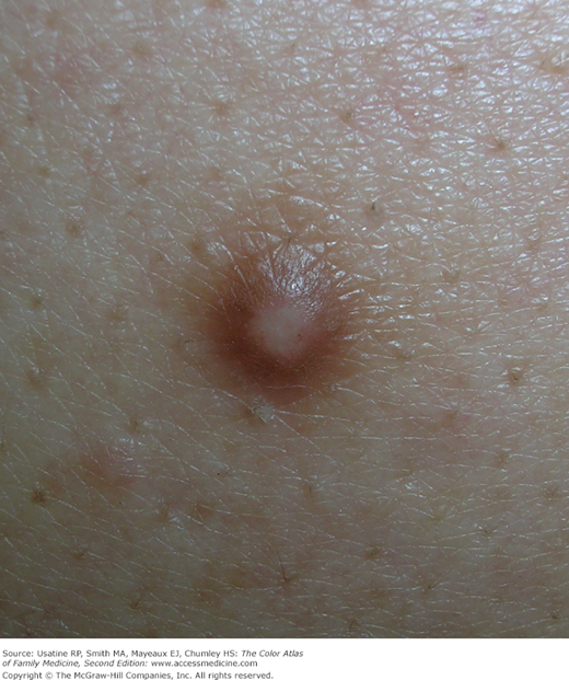
A 25-year-old woman reports a firm nodule on her leg that gets in the way of shaving her leg (Figure 160-1). Upon questioning, the nodule may have started there after she cut her leg shaving 1 year ago. She is worried it could be a cancer and wants it removed. Close observation showed a brown halo and a firm nodule that dimpled down when pinched. A diagnosis of a dermatofibroma (DF) was made and the choices for treatment were discussed.
Introduction
Epidemiology
Etiology and Pathophysiology
- Uncertain etiology—Nodule may represent a fibrous reaction triggered by trauma, a viral infection, or insect bite; however, DFs show clonal proliferative growth seen in both neoplastic and inflammatory conditions.3
- Multiple DFs (i.e., >15 lesions) have been reported associated with systemic lupus erythematosus, HIV infection, Down syndrome, Graves disease, or leukemia, and may represent a worsening of immune function.1 A case of familial eruptive DFs has also been reported associated with atopic dermatitis.4
Diagnosis
- Firm to hard nodule; skin is freely movable over the nodule, except for the area of dimpling.
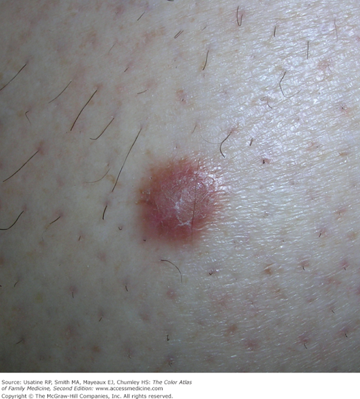
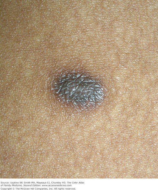
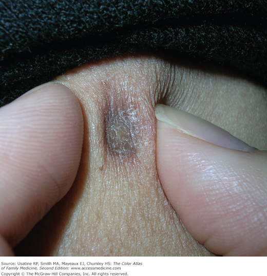
- Color of the overlying skin ranges from flesh to gray, pink, red, blue, brown, or black (Figures 160-2 and 160-3), or a combination of hues (Figure 160-4).
- Dimples downward when compressed laterally because of tethering of the overlying epidermis to the underlying nodule (Figure 160-3).
- Usually asymptomatic but may be tender or pruritic.
- Size ranges from 0.3 to 10 mm; usually less than 6 mm. Rarely, DFs grow to larger than 5 cm.5
- May have a hyperpigmented halo and a scaling surface (Figure 160-4).
- DFs can rarely be located entirely within subcutaneous tissue.6
- Dermoscopy is a useful adjunctive diagnostic technique for DF (Figure 160-5). Although the most common finding is a peripheral pigment network with a central white area (34.7% of cases), 10 dermoscopic patterns have been identified; in a large case series, pigment network was observed in 71.8% (3% atypical pigment network).7 (See Appendix C: Dermoscopy.)