Dermal Nerve Sheath Myxoma

 Pretibial skin also common


image Uncommon on face


• Benign but up to 50% recur

• Rare intraoral, orbital, intradural, and paraspinal cases


Microscopic


• Multiple lobules with abundant myxoid matrix

• Well-defined lobular architecture and dense fibrous septa

• Usually limited to dermis and subcutis

• Spindle-shaped and epithelioid cells

• Interconnecting cords, networks, syncytial nests, ring-like structures

• Stellate and vacuolated cells common

• No atypia, very low mitotic rate


Ancillary Tests


• Diffuse S100(+)

• GFAP(+) in most

• SMA, desmin, CD68, HMB-45, synaptophysin, chromogranin-A, CD31 (-)


Top Differential Diagnoses


• Neurothekeoma

• Superficial angiomyxoma

• Digital mucoid cyst

• Myxoid neurofibroma

• Superficial acral fibromyxoma

• Myxoid dermatofibrosarcoma protuberans

• Myxofibrosarcoma (low grade)

image
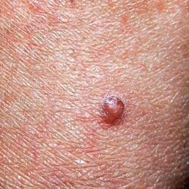
Dermal Papule
Dermal nerve sheath myxoma typically presents as a firm papule that varies from 0.5-4.5 cm in size. It usually occurs in adults. Common locations are the finger, hand, and pretibial skin.


image
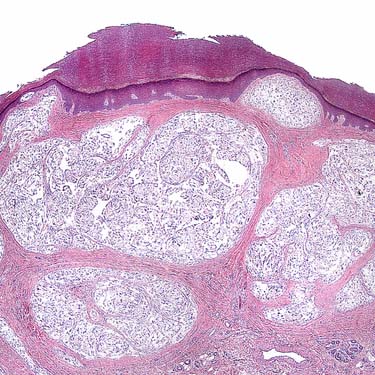
Dermal Nerve Sheath Myxoma
Dermal nerve sheath myxoma has a very distinctive architecture, consisting of well-defined myxoid lobules. It is typically centered within the dermis and can involve the underlying subcutis as well.

image
Composed of Schwann Cells
The cells in dermal nerve sheath myxoma have schwannian differentiation. They have uniform spindle-shaped or oval nuclei, evenly distributed chromatin, and long fibrillary cytoplasmic processes image 
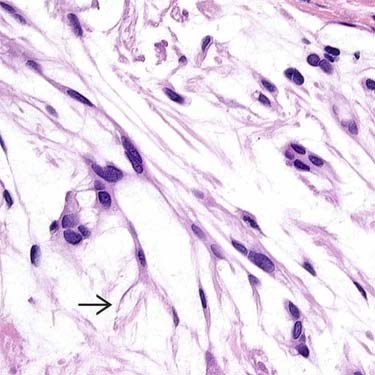
forming bipolar and stellate configurations.
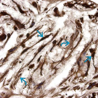
image
Strong, Diffuse S100 Expression
Dermal nerve sheath myxomas are strongly S100(+) consonant with Schwann cell differentiation. This immunohistochemically stained section depicts diffuse and intense nuclear image and cytoplasmic image reactivity.


TERMINOLOGY


Synonyms


• Cutaneous lobular neuromyxoma, perineurial myxoma, pacinian neurofibroma, myxoid tumor of nerve sheath


Definitions


• Benign cutaneous nerve sheath tumor with Schwann cell differentiation, abundant myxoid matrix, and well-defined lobular architecture


CLINICAL ISSUES


Epidemiology


• Incidence
image Rare; fewer than 100 reports

• Age
image Mostly adults; median age: 34 years


Site


• Extremities
image Fingers and hand most common

image Pretibial skin common

• Rare intraoral, orbital, intradural, and paraspinal cases

• Uncommon on face


Presentation


• Slowly growing, painless mass


Treatment


• Surgical approaches
image Simple excision


Prognosis


• Benign and nonaggressive but often excised with positive margins
image Up to 50% recur


MACROSCOPIC


Size


• 0.5-4.5 cm


MICROSCOPIC


Histologic Features


• Multiple myxoid lobules
image Well-defined borders separated by dense fibrous septa

• Usually limited to dermis and subcutis

• Paucicellular proliferation of bland spindle cells
image Interconnecting cords, networks, syncytial nests, ring-like structures, Verocay-like bodies

Only gold members can continue reading. Log In or Register to continue

Stay updated, free articles. Join our Telegram channel

Apr 24, 2017 | Posted by in PATHOLOGY & LABORATORY MEDICINE | Comments Off on Dermal Nerve Sheath Myxoma

Full access? Get Clinical Tree

Get Clinical Tree app for offline access