Dermal Melanocytoses (Nevus of Ota and Ito, Mongolian Spot)
Christine J. Ko, MD
Key Facts
Clinical Issues
Nevus of Ota and Ito and Mongolian spot
Often evident at birth
Occasionally manifest later in life
Generally stable over time (nevus of Ota and Ito); resolution with time (Mongolian spot)
Blue to gray macule/patch
Nevus of Ota: Near/involving eye
Nevus of Ito: Shoulder area
Mongolian spot: Generally on back
Microscopic Pathology
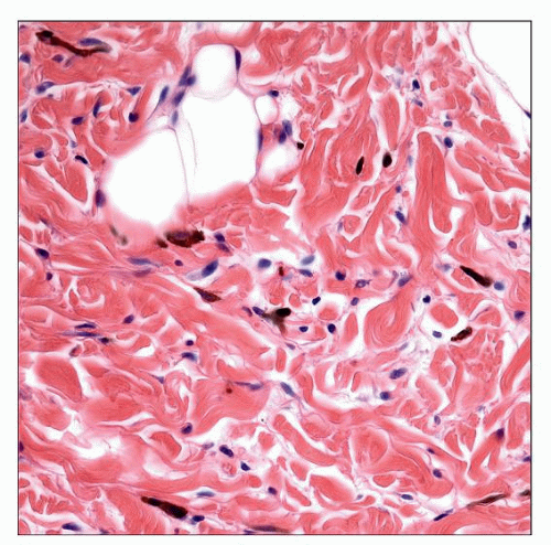
Dendritic melanocytes in dermis
Dermis generally otherwise normal (without sclerosis)
Top Differential Diagnoses
Blue nevus
Hori nevus
Becker nevus
Child abuse
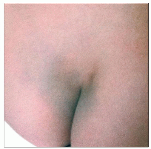
 This is the typical location for a Mongolian spot, over the sacral area. There may also be patches in other areas of the back. |
CLINICAL ISSUES
Epidemiology
Age
Often evident at birth, but occasionally manifest later
Ethnicity
More common in Asians
Site
Nevus of Ota
Located near/involving the eye (conjunctiva)
In distribution of trigeminal nerve (ophthalmic/maxillary)
Nevus of Ito
Located on shoulder/deltoid area
Mongolian spot
Located most commonly on the back, especially lumbosacral
Rarely, in so-called “aberrant” form, may involve extremities
When located in areas other than the back, overlaps with blue nevi
Presentation
Nevus of Ota/Ito
Often unilateral
Color often blue to gray
Variably sized mottled patch
Mongolian spot
Generally 1 or more patches of blue to gray
Stay updated, free articles. Join our Telegram channel

Full access? Get Clinical Tree


