Embryonal, alveolar, pleomorphic, and epithelioid types of RMS have been described as primary cutaneous tumors
Clinical Issues
• Very rare
Cutaneous metastases or secondary involvement of skin by deep primary must be thoroughly excluded
• Bimodal age distribution
Children and young adults: Embryonal and alveolar types
Older adults: Plemorphic and epithelioid types
• May occur at any cutaneous site
• Limited data available regarding treatment and prognosis
However, recurrences and metastases reported
Microscopic
• Confined to dermis &/or subcutaneous tissue
• Embryonal rhabdomyosarcoma (RMS)
Variably cellular sheets of spindle and ovoid cells
Myxoid stroma common
• Alveolar RMS
Nests of small round cells with fibrous septa
Multinucleated tumor giant cells (“wreath cells”) may be present
• Pleomorphic RMS
Sheets of markedly pleomorphic, atypical cells
May show rhabdoid morphology
• Epithelioid RMS
Sheets of uniform epithelioid cells with abundant eosinophilic cytoplasm
Eccentric nuclei with prominent nucleoli
Ancillary Tests
• Desmin (+), myogenin (+), MYOD1(+)
• Focal keratin (+) reported
• Negative for S100, SOX10, p63, CD31, melanocytic markers
Top Differential Diagnoses
• Superficial Ewing sarcoma/PNET
• Carcinoma, melanoma
• Merkel cell carcinoma
• Atypical fibroxanthoma
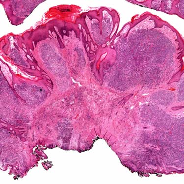
Cutaneous Rhabdomyosarcoma This cutaneous rhabdomyosarcoma (RMS) from the ear is a polypoid tumor with extensive infiltration of the dermis by hypercellular sheets of fairly uniform, small, hyperchromatic, ovoid cells.
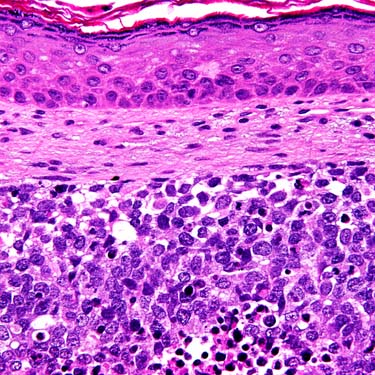
Cutaneous Rhabdomyosarcoma at High Power High magnification of a cutaneous RMS shows a nodular dermal proliferation of medium-sized, blue-staining, ovoid cells. A grenz zone separates the tumor from overlying epidermis.
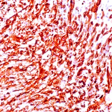
Desmin Expression in Rhabdomyosarcoma Desmin expression in RMS is typically diffuse and strong in the cytoplasm of the neoplastic cells. Cross striations may be highlighted.
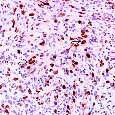
Myogenin Expression in Rhabdomyosarcoma Immunostaining for myogenin shows well-defined nuclear localization . The proportion of nuclei expressing this marker varies. It is most widespread in alveolar RMS, with lesser degrees in embryonal and pleomorphic subtypes. Cytoplasmic staining is nonspecific and seen in many tumor types.
TERMINOLOGY
Abbreviations
• Rhabdomyosarcoma (RMS)
Definitions
• Malignant mesenchymal neoplasm arising in dermis &/or subcutis and showing variable differentiation toward skeletal muscle
Cutaneous metastases or secondary involvement by deep primary tumor must be excluded
Embryonal, alveolar, pleomorphic, and epithelioid types of RMS have been described as primary cutaneous tumors
CLINICAL ISSUES
Epidemiology
• Incidence
However, primary cutaneous RMS is very rare
– Cutaneous metastases or secondary involvement of skin must be thoroughly excluded
• Age
Bimodal age distribution
– Children and young adults
Embryonal and alveolar types most common
– Older adults
Pleomorphic and epithelioid types most common
• Sex
M = F
Site
• May occur at any cutaneous site
Including head and neck, trunk, extremities
Presentation
• Often presents as asymptomatic papule or nodule
May cause pain
• Nonspecific clinical features
Diagnosis may therefore be delayed
Treatment
• Complete surgical excision
• Chemotherapy &/or radiation
Prognosis
• Data limited due to small number of cases reported
• However, documented cutaneous cases prone to repeated recurrences and metastases (including to lymph nodes and lung)
• Overall mortality rate 36% in one small series
MACROSCOPIC
Size
• Usually 1-4 cm
MICROSCOPIC
Histologic Features
• May be confined to dermis, subcutis, or involve both dermis and subcutis
• May show ulceration of overlying skin
• Rhabdomyoblasts
Cells with eccentric nuclei and variable amounts of eosinophilic cytoplasm
Cytoplasmic cross striations may be visible
Variable numbers and stages of differentiation
Can be found in all RMS subtypes
Only gold members can continue reading. Log In or Register to continue


 . The proportion of nuclei expressing this marker varies. It is most widespread in alveolar RMS, with lesser degrees in embryonal and pleomorphic subtypes. Cytoplasmic staining is nonspecific and seen in many tumor types.


