Conjunctival Melanoma Presenting as Pigmented Patch Clinical image shows an expansile pigmented patch at the limbus and adjacent to the iris . Bulbar conjunctiva close to the limbus is the most common location for primary acquired melanosis (PAM).
Scanning Magnification of PAM With Atypia Scanning magnification of this conjunctival shave biopsy shows a lentiginous basilar proliferation that stands out even at the this power. The radial extension appears to be extensive.
Higher Magnification of PAM With Atypia Higher magnification of the same case confirms that atypical melanocytes have replaced the basilar keratinocytes in the conjunctiva. There is some pagetoid upward scatter of melanocytes .
Higher Magnification Shows Nuclear Hyperchromasia and Pleomorphism High-power magnification shows poorly nested proliferation of atypical melanocytes in the same lesion of PAM with atypia. The upward scatter , nuclear hyperchromasia , and nuclear pleomorphism resembles cutaneous melanoma in situ.
TERMINOLOGY
Abbreviations
• Conjunctival melanoma (CM)
Synonyms
• Conjunctival malignant melanoma
Definitions
• Malignant proliferation of conjunctival melanocytes commonly associated with primary acquired melanosis (PAM) with atypia
CLINICAL ISSUES
Epidemiology
• Incidence
Rare; accounts for only 2-3% of ocular cancers
Accounts for 1% of noncutaneous malignant melanoma
• Age
More common in older individuals (mean age at presentation: 50-60 years)
Exceedingly rare in children younger than 15 years of age (only 28 reported cases)
Presentation
• Asymptomatic raised, pigmented plaque, macule, or tumor
• Can range in size from millimeters to large tumor masses
• Bulbar conjunctiva close to limbus is most common location
Natural History
• Majority of cases (53-75%) arise in setting of PAM with atypia
• Remainder (18-30%) arise de novo
• Minority of cases (5%) are associated with preexisting melanocytic nevus
Treatment
• Options, risks, complications
Avoiding manipulation of tumor during surgery reduces local recurrence rate and lymphatic spread
• Surgical approaches
All should be completely excised with 2- to 3-mm tumor-free margins
Sentinel lymph node biopsy recommended for high-risk tumors
– > 10 mm in diameter and > 2 mm in thickness
– Nonlimbus locations
• Adjuvant therapy
Risk of recurrence is reduced with adjuvant cryotherapy, irradiation, or topical chemotherapy
Prognosis
• Conjunctival melanoma arising in association with PAM has higher risk of local recurrence
• 5-year survival rate: 87-95%
• 10-year survival rate: 70-86%
• After 10 years, 50% of tumors will recur locally and about 25% will metastasize
• Adverse clinical prognostic indicators include
Nonbulbar (fornix, palpebral) location
Involvement of lymphatics-rich caruncle
Only gold members can continue reading. Log In or Register to continue
 at the limbus
  and adjacent to the iris
  . Bulbar conjunctiva close to the limbus is the most common location for primary acquired melanosis (PAM).
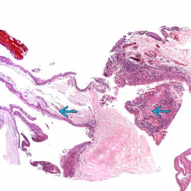
 that stands out even at the this power. The radial extension appears to be extensive.
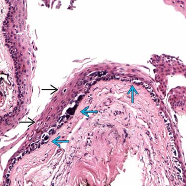
 have replaced the basilar keratinocytes in the conjunctiva. There is some pagetoid upward scatter of melanocytes
  .
.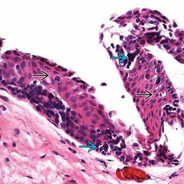
 , nuclear hyperchromasia
  , and nuclear pleomorphism resembles cutaneous melanoma in situ.


