Conjunctival melanocytic nevi (50%) can exhibit numerous epithelial-lined cysts  located within the lesion, as seen here under scanning magnification. This is a useful finding, supporting benignancy. Note the lymphohistiocytic infiltrate
  hugging the lesion.
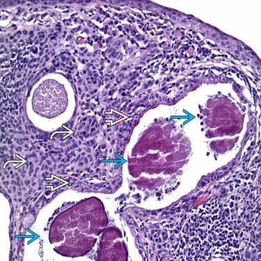
Higher power magnification shows the cysts to contain sulfur granules
 and invoke a neutrophilic response
  . The nested nevomelanocytes
  , surrounding the infected cysts, are devoid any cytological atypia.
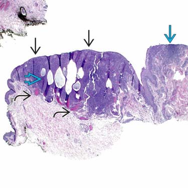
At this scanning magnification, a hypercellular proliferation appears to occupy the subepithelium
 underneath the conjunctival epithelium
  . There is intralesional cystic degeneration
  . There is a focus of intense lymphohistiocytic infiltrate
  commonly seen in these lesions.
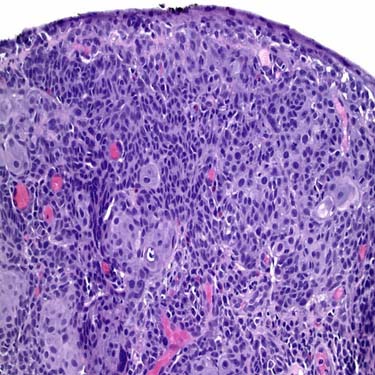
A higher magnification is shown from the same lesion. The subepithelial component does not appear to decrease in the nuclear:cytoplasmic ratio. This paradoxical maturation is an expected feature.


MICROSCOPIC
Histologic Features
• Anatomic classification (similar to skin)
 Junctional: Nested but sometimes also lentiginous proliferations of type A or type B cells confined to epithelium


Junctional: Nested but sometimes also lentiginous proliferations of type A or type B cells confined to epitheliumStay updated, free articles. Join our Telegram channel
Full access? Get Clinical Tree
