• Lesion overall orderly, symmetric, and well circumscribed
• Most commonly: Compound/intradermal melanocytic nevus plus blue nevus
• Others types of combined nevi include compound or intradermal nevus plus pigmented spindle or spitzoid cells
Top Differential Diagnoses
• Blue nevus (common and cellular types)
Composed of dendritic to spindled melanocytes and melanophages
Cellular often shows deep extension into dermis/subcutaneous
• Deep penetrating nevus
Wedge-shaped architecture
Often deep extension into dermis/subcutaneous
Junctional nests may be present
Dermal component composed of epithelioid/spindled melanocytes in nests bordered by melanophages
Nests/fascicles of cells may be centered around adnexal/neurovascular structures in dermis
Occasionally, bulbous/pushing margin is present
• Congenital melanocytic nevus: Present at birth
May extend deep into dermis or subcutaneous tissue
May infiltrate arrector pili, adnexal structures, nerves
Maturation with depth
Combined Nevus With Conventional and Blue Nevus Components This is an example of a combined nevus of the most common type (i.e., composed of a conventional compound melanocytic nevus and a blue nevus ). The lesion is symmetric and bland appearing.
Combined Nevus: Conventional and Spitz This combined nevus has conventional nevomelanocytes (small cells with oval to round nuclei, some in round nests) as well as larger cells with eosinophilic cytoplasm and larger nuclei (spitzoid cells ).
Differential Diagnosis: Spitz Nevus, Desmoplastic Type The differential diagnosis of a combined nevus may include other less common nevi, such as a desmoplastic Spitz nevus. The cells are large with eosinophilic cytoplasm . There are interspersed melanophages and a sclerotic stroma .
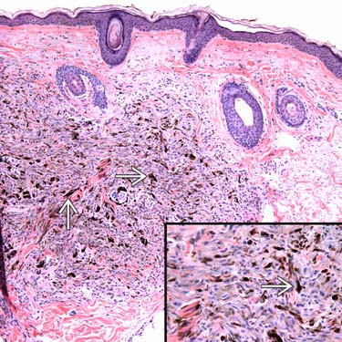
Differential Diagnosis: Deep Penetrating Nevus The differential diagnosis of a combined nevus can also include a deep penetrating nevus, shown here, with a nodular collection of finely pigmented spindled to epithelioid cells in fascicles with many interspersed melanophages .
TERMINOLOGY
Synonyms
• Melanocytic nevus with phenotypic heterogeneity, clonal nevus, nevus with focal epithelioid component, combined Spitz nevus, inverted type A nevus
Definitions
• Presence of 2 or more distinct populations of melanocytes (i.e., type A melanocytic nevus cells and spindled or dendritic cells) or
• Presence of 2 or more types of melanocytic nevi (i.e., intradermal melanocytic and blue or Spitz)
CLINICAL ISSUES
Presentation
• Generally in young adults
Only gold members can continue reading. Log In or Register to continue


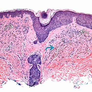
 and a blue nevus
  ). The lesion is symmetric and bland appearing.
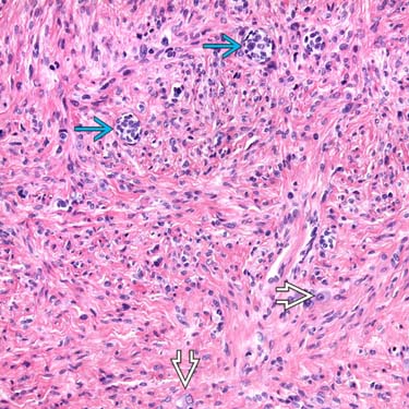
 (small cells with oval to round nuclei, some in round nests) as well as larger cells with eosinophilic cytoplasm and larger nuclei (spitzoid cells
  ).
).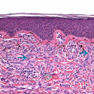
 . There are interspersed melanophages and a sclerotic stroma
  .
.
 .
.


