Chapter 29 Colposcopy Examination
Common indications
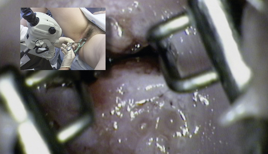
Colposcopy is used to evaluate cervical abnormalities found on Pap smear cytology, visual screening with acetic acid, or to evaluate visible abnormalities (Figure 29-1).
Equipment
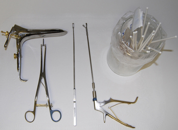
The instruments needed for this procedure include an adjustable vaginal speculum, an endocervical speculum, an endocervical curette, biopsy forceps, and small and large cotton-tipped swabs. A binocular colposcope provides optimal visualization and should have a green light filter. Use Monsel’s solution for chemical cautery after biopsy (Figure 29-2).
Key steps
1. Preparation: Place the patient in the lithotomy position, and insert a lubricated speculum. Clearly visualize the cervix, and focus the colposcope. Note any obvious abnormalities, and identify the transformation zone, the os, and the squamocolumnar junction (Figure 29-3).
2. Acetic acid application: Apply 5% acetic acid to the cervix with two large swabs. Acetic acid converts potentially dysplastic epithelium to a whitish color within about 30 seconds. The cervix is then carefully inspected to look for any distinctive changes consistent with dysplasia. Acetowhite changes may take longer to develop in areas of high-grade dysplasia. Identify areas of fine mosaicism, coarse mosaicism, or punctuation. Green light examination allows for better visualization of a cervical vascular pattern (Figure 29-4A, B).
3. Endocervical examination: Examine the endocervical canal using an endocervical speculum to open the distal endocervical canal. The glandular endocervical epithelium can become faintly white; however, dysplasia appears more plaque-like and distinctly more acetowhite. Identifying lesions that lie within or extend into the endocervical canal is a critical requirement for a satisfactory colposcopy examination (Figure 29-5).
4. Lugol’s application: Concentrated Lugol’s iodine solution is also a useful adjunct in highlighting abnormal areas in patients without iodine allergy. Saturate swab, then paint entire surface. Dry cervix lightly after staining to remove residual Lugol’s solution. Areas that stain dark brown are normal squamous epithelium, whereas acetowhite areas that stain yellow are more likely to be low-grade dysplasia. Areas that remain unstained despite application are more likely to be highly dysplastic tissue if they are within an area that was already abnormal with the acetowhite examination (Figure 29-6).
Stay updated, free articles. Join our Telegram channel

Full access? Get Clinical Tree


