6 Clinical biochemistry and metabolism
Between 60 and 70% of all critical decisions taken in regard to patients in health-care systems in developed countries involve a laboratory service or result. This chapter describes disorders whose primary manifestation is in abnormalities of biochemistry laboratory results, or whose underlying pathophysiology involves disturbance in specific biochemical pathways. Discussion of diabetes mellitus and other endocrine disorders is to be found in Chapters 10 and 11.
WATER AND ELECTROLYTE DISTRIBUTION
In a typical adult male, the 40 litres of total body water (TBW) constitute ∼60% of the body weight. More than half is located inside cells (the intracellular fluid or ICF), while the remainder is in the extracellular fluid (ECF) compartment. Of the ECF, the plasma is itself a small fraction (some 3 litres), while the remainder is interstitial fluid within the tissues but outside the cells.
The dominant cation in the ICF is potassium, while in the ECF it is sodium (Fig. 6.1). Phosphates and negatively charged proteins constitute the major intracellular anions, while chloride and, to a lesser extent, bicarbonate dominate the ECF anions. An important difference between the plasma and interstitial ECF is that only plasma contains significant concentrations of protein.

Fig. 6.1 Normal distribution of body water and electrolytes. Schematic representation of volume (l = litres) and composition (dominant ionic species only shown) of the intracellular fluid (ICF) and extracellular fluid (ECF) in a 70 kg male. Total body water (TBW) = ∼60% of body weight. TBW = ICF + ECF = 40 litres. The main difference in composition between the plasma and interstitial fluid (ISF) is the presence of appreciable concentrations of protein in the plasma (not shown) but not the ISF. The sodium–potassium pump maintains the cation concentration difference between the ICF and ECF. The difference in protein content between the plasma and interstitial compartments is maintained by the permeability characteristics of the capillary wall.
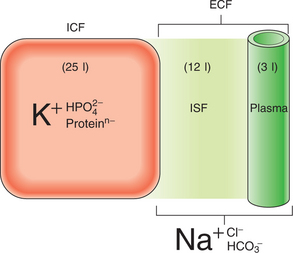
The major force maintaining the difference in cation concentration between the ICF and ECF is the sodium–potassium pump (Na,K-activated ATPase) integral to all cell membranes. Maintenance of the cation gradients across cell membranes is essential for many cell processes, including the excitability of conducting tissues such as nerve and muscle. The difference in protein content between the plasma and the interstitial fluid compartment is maintained by the protein permeability barrier at the capillary wall. This protein concentration gradient contributes to the balance of forces across the capillary wall favouring fluid retention within the capillaries (the colloid osmotic, or oncotic, pressure of the plasma), thus maintaining an adequate circulating plasma volume.
INVESTIGATION OF WATER AND ELECTROLYTES
Because the blood consists of both intracellular (red cell) and extracellular (plasma) components, it is important to avoid haemolysis of the sample, which causes contamination of the plasma by intracellular elements, particularly potassium. Blood should not be drawn from an arm into which an i.v. infusion is being given, to avoid contamination by the infused fluid.
Since the kidney maintains body fluid composition by adjusting urine volume and composition, it is often helpful to obtain a simultaneous sample of urine (‘spot’ specimen or 24-hr collection) at the time of blood analysis.
DISORDERS OF SODIUM BALANCE
When sodium balance is disturbed, as a result of imbalance between intake and excretion, any tendency for plasma sodium concentration to change is usually corrected by the osmotic mechanisms controlling water balance (see below). As a result, disorders in sodium balance present chiefly as altered ECF volume rather than altered sodium concentration.
SODIUM DEPLETION (USUALLY ASSOCIATED WITH HYPOVOLAEMIA)
Aetiology includes the following factors:
Clinical features
Symptoms and signs of hypovolaemia are as follows:
Supportive biochemistry includes:
Management of sodium and water depletion
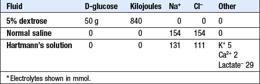
I.v. fluid therapy: Box 6.1 shows the daily maintenance requirements for water and electrolytes in a typical adult, and Box 6.2 summarises the composition of some widely available i.v. fluids. The choice of fluid and the rate of administration will depend on the clinical circumstances, as assessed at the bedside and from laboratory data.
6.1 BASIC DAILY WATER AND ELECTROLYTE REQUIREMENTS
Requirement per kg | Typical 70 kg adult | |
---|---|---|
Water | 35–45 ml/kg | 2.5–3.0 l/day |
Sodium | 1.5–2 mmol/kg | 100–140 mmol/day |
Potassium | 1.0–1.5 mmol/kg | 70–100 mmol/day |
NB for Na and K, mmol = meq
SODIUM EXCESS (USUALLY ASSOCIATED WITH HYPERVOLAEMIA)
In the presence of normal function of the heart and kidneys, an excessive intake of salt and water is compensated for by increased excretion and so is unlikely to lead to clinically obvious features of hypervolaemia.
Causes of sodium and water excess in clinical practice include:
Peripheral oedema is the most common physical sign associated with these conditions (p. 174) although it is not usually a feature of Conn’s syndrome.
Management
The management of ECF volume overload involves:
DISORDERS OF WATER BALANCE
Daily water intake can vary over a wide range, from 500 ml to several litres a day. While a certain amount of water is lost through the stool, sweat and the respiratory tract, the kidneys are chiefly responsible for adjusting water excretion to maintain a constant body water content and body fluid osmolality (normal range 280–300 mmol/kg).
Disturbances in body water metabolism, in the absence of changes in sodium balance, manifest principally as abnormalities of plasma sodium concentration, and hence of plasma osmolality. The main consequence of changes in plasma osmolality, especially when rapid, is altered cerebral function. This is because, when extracellular osmolality changes abruptly, water flows rapidly across cell membranes with resultant cell swelling (during hypo-osmolality) or shrinkage (during hyperosmolality). Cerebral cell function is very sensitive to such volume changes, particularly during cell swelling where an increase in intracerebral pressure occurs due to the constraints posed by the bony skull, resulting in impaired cerebral perfusion.
HYPONATRAEMIA
The causes of hyponatraemia (plasma Na <135 mmol/l) are best organised according to any associated change in ECF volume status, i.e. the total body sodium. In all cases, there is retention of water relative to sodium, and it is the clinical examination rather than the electrolyte test results which gives clues to the underlying problem.
Hypovolaemic (sodium deficit with a relatively smaller water deficit): Renal Na loss (diuretics), GI Na loss (vomiting, diarrhoea).
Euvolaemic (water retention alone, i.e. ‘dilutional’): Primary polydipsia, SIADH (Box 6.3).
6.3 SYNDROME OF INAPPROPRIATE ANTIDIURETIC HORMONE SECRETION (SIADH): CAUSES AND DIAGNOSIS
Hypervolaemic (sodium retention with relatively greater water retention): Heart failure, cirrhosis, chronic kidney disease (without water restriction).
Clinical features
When hyponatraemia develops gradually, it is relatively asymptomatic. More rapid changes in plasma osmolality and so plasma sodium may be associated with:
Investigations
Plasma and urine electrolytes and osmolality (Box 6.4) are usually the only tests required to classify the hyponatraemia.
6.4 URINE Na AND OSMOLALITY IN THE DIFFERENTIAL DIAGNOSIS OF HYPONATRAEMIA*
Urine Na (mmol/l) | Urine osmolality (mmol/kg) | Possible diagnoses |
---|---|---|
Low (<30) | Low (<100) | Primary polydipsia, malnutrition |
Low | High (>150) | Salt depletion, hypovolaemia |
High (>40) | Low | Diuretic action (acute phase) |
High | High | SIADH |
* Note that intermediate urine results are of indeterminate significance and diagnosis depends on a comprehensive clinical assessment.
Management
The treatment for hyponatraemia is critically dependent on the rate of development and severity, and on the underlying cause.
In general, if hyponatraemia has developed rapidly (over hours to days), morbidity will be high due to cerebral oedema, and it is generally safe to correct the plasma sodium relatively rapidly. This can include infusion of hypertonic (3%) sodium chloride solutions, especially when the patient is obtunded or convulsing.
Rapid correction of hyponatraemia that has developed slowly (over weeks to months) may lead to ‘central pontine myelinolysis’, which may cause permanent structural and functional cerebral changes and is generally fatal. The rate of plasma sodium correction in chronic asymptomatic hyponatraemia should not exceed 10 mmol/l/day, and an even slower rate would generally be safer.
Specific treatment should be directed at the underlying cause.
HYPERNATRAEMIA
Just as hyponatraemia represents a failure of the mechanisms for diluting the urine during free access to water, so hypernatraemia (plasma Na > 150 mmol/l) reflects an inadequacy of the kidney in concentrating the urine in the face of relatively restricted water intake. Similar to hyponatraemia, the causes of hypernatraemia may be classified based on the volume state of the patient.
Hypovolaemic (sodium deficit with a relatively greater water deficit): Renal Na losses (diuretics), GI Na losses (colonic diarrhoea), skin Na losses (excessive sweating).
Euvolaemic (water deficit alone): Diabetes insipidus (central or nephrogenic, p. 377).
Clinical features
Patients with hypernatraemia generally have reduced cerebral function and cerebral dehydration. This triggers thirst and drinking, and if adequate water is obtained, is self-limiting. If adequate water is not obtained, dizziness, confusion, weakness and ultimately coma and death can result.
Management
Treatment of hypernatraemia depends on both the rate of development and the underlying cause.
If there is reason to think that the condition has developed rapidly, correction with appropriate volumes of i.v. hypotonic fluid may be attempted relatively rapidly.
In older institutionalised patients however, it is more likely that the disorder has developed slowly and extreme caution should be exhibited in lowering the plasma sodium rapidly, to avoid the risk of cerebral oedema.

Stay updated, free articles. Join our Telegram channel

Full access? Get Clinical Tree

