Patient Story
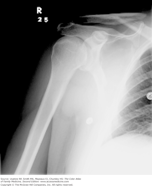
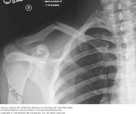
A 17-year-old boy presents after falling off his skateboard and landing directly on his lateral shoulder. He had immediate pain and swelling in the middle of his clavicle. His examination revealed a bump in the middle of his clavicle. A radiograph confirmed a midclavicular fracture (Figure 102-1). He was treated conservatively with a sling, which he wore for approximately 1 of the recommended 3 weeks. A follow-up radiograph demonstrated good healing. The bump on his clavicle is still palpable; however, this does not bother him.
Figure 102-1
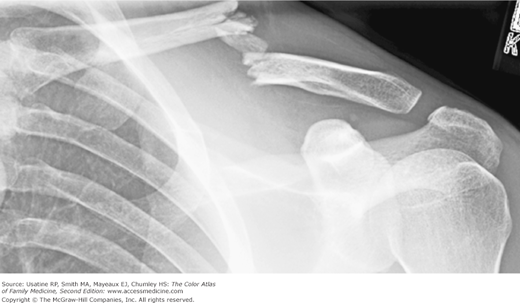
Midshaft clavicle fracture. Clavicle fractures are designated midshaft (in the middle third), distal (distal third), or medial (medial third). (From Simon RR, Sherman SC, Koenigsknecht SJ. Emergency Orthopedics the Extremities. New York, NY: McGraw-Hill; 2007:286, Fig. 11-35. Copyright 2007.)
Introduction
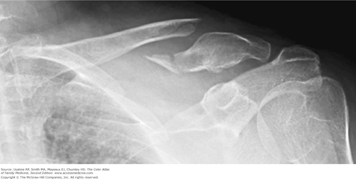
Clavicular fractures are common in both children and adults and are most commonly caused by accidental trauma. The clavicle most commonly fractures in the midshaft (Figures 102-1, 102-2, 102-3), but can also fracture distally (Figure 102-4). Many fractures can be treated conservatively. Refer patients with significant displacement or distal fractures for surgical evaluation.
Epidemiology
Etiology and Pathophysiology
- Most are caused by accidental trauma from fall against the lateral shoulder or an outstretched hand or direct blow to the clavicle; however, stress fractures in gymnasts and divers have been reported.
- Pathologic fractures (uncommon) can result from lytic lesions, bony cancers or metastases, or radiation.
- Birth trauma (neonatal).
- Physical assaults, intimate partner violence, and child abuse can cause clavicular fractures.
Diagnosis
- History of trauma with a mechanism known to result in clavicle fractures (i.e., fall on an outstretched hand or lateral shoulder, or direct blow).
- Pain and swelling at the fracture site.
- Gross deformity at site of fracture.
Stay updated, free articles. Join our Telegram channel

Full access? Get Clinical Tree


