Patient Story
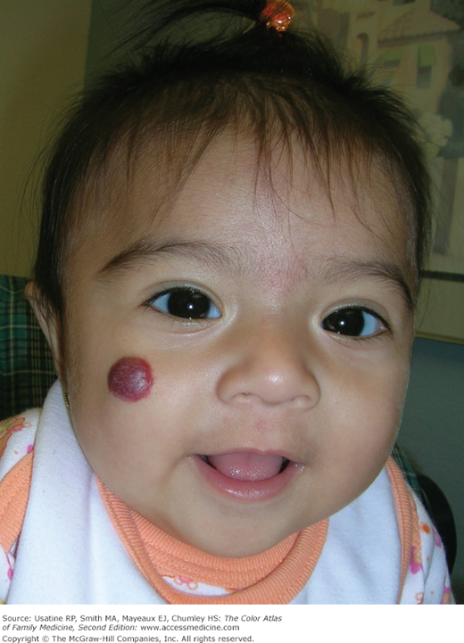
A baby girl is brought to the office because her mother is concerned over the growing strawberry hemangioma on her face. Her mother is reassured that most of these childhood hemangiomas regress over time and that there is no need for immediate treatment (Figure 109-1).
Introduction
Synonyms
Epidemiology
- Approximately 30% of hemangiomas are present at birth; the other 70% appear within the first few weeks of life.
- Hemangiomas occur more commonly in fair-skinned, premature, female infants. In one study, the mothers of children with hemangiomas are of higher maternal age, have a higher incidence of preeclampsia and placenta previa, and are more likely to have had multiple gestation pregnancies.1
- There is an increased incidence of vascular anomalies in the families of children born with hemangiomas.1
- The data are mixed as to whether chorionic villus sampling may play a role in the formation of hemangiomas.1
- Females are affected more often than males (2.4:1).1
Etiology and Pathophysiology
- Hemangiomas consist of an abnormally dense group of dilated blood vessels. Most childhood hemangiomas are thought to occur sporadically.
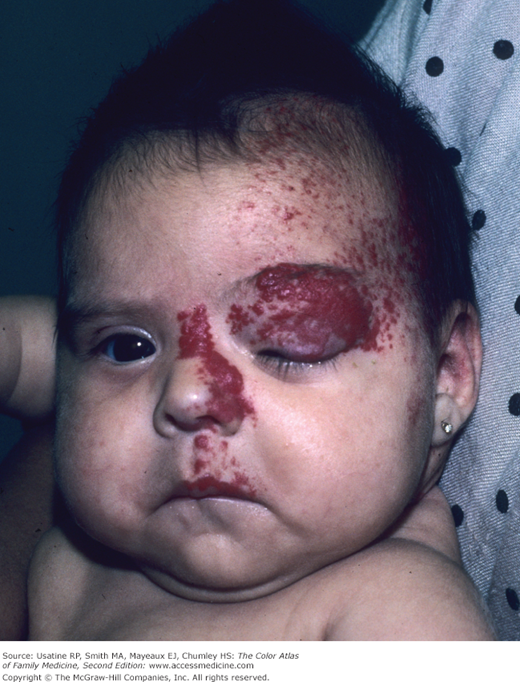
- Hemangiomas are characterized by an initial phase of rapid proliferation, followed by spontaneous and slow involution, often leading to complete regression. Most childhood hemangiomas are small and innocuous, but some grow to threaten a particular function (Figure 109-2) or even life.
- Rapid growth during the first month of life is the historical hallmark of hemangiomas, when rapidly dividing endothelial cells are responsible for the enlargement of these lesions. The hemangiomas become elevated and may take on numerous morphologies (dome-shaped, lobulated, plaque-like, and/or tumoral). The proliferation phase occurs during the first year, with most growth taking place during the first 6 months of life. Proliferation then slows and the hemangioma begins to involute.
- The involutional phase may be rapid or prolonged. No specific feature has been identified in explaining the rate or completeness of involution. However, in one type of hemangioma, the rapidly involuting congenital hemangioma, the proliferation phase occurs entirely in utero such that the lesion is fully developed at birth, followed by complete involution during the second year of life.1
- A good rule of thumb is 50% of childhood hemangiomas will involute by age 5 years, 70% by age 7 years, and the remainder of childhood hemangiomas will take an additional 3 to 5 years to complete the process of involution.1
- Of the lesions that have involuted by age 6 years, 38% will leave residual evidence of the hemangioma in the form of a scar, telangiectasia, or redundant, “bag-like” skin. The chance of a permanent scar increases the longer it takes to involute. For example, of the lesions that involute after age 6 years, 80% may exhibit a cosmetic deformity.1
Diagnosis
Early lesions may be subtle, resembling a scratch or bruise, or alternatively may look like a small patch of telangiectasias or an area of hypopigmentation. Hemangiomas can start off as a flat red mark, but as proliferation ensues, it grows to become a spongy mass protruding from the skin. The earliest sign of a hemangioma is blanching of the involved skin with a few fine telangiectasias followed by a red macule. Rarely, a shallow ulceration may be the first sign of an incipient hemangioma.1 Hemangiomas are typically diagnosed based on appearance, rarely warranting further diagnostic tests.
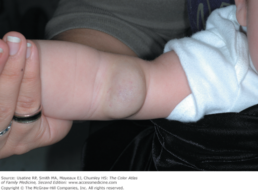
Hemangiomas may be superficial, deep, or a combination of both. Superficial hemangiomas are well defined, bright red, and appear as nodules or plaques located above clinically normal skin (Figures 109-1, 109-2, 109-3). Deep hemangiomas are raised flesh-colored nodules, which often have a bluish hue and feel firm and rubbery (Figure 109-4).
Figure 109-3
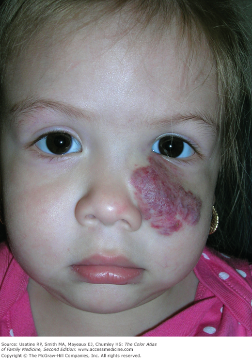
Strawberry hemangioma present since birth on the face of a 22-month-old girl. Although it is close to her eye, her vision has never been occluded. She has been followed by ophthalmology and no active treatment was recommended. The hemangioma grew larger during the first year of life and is now beginning to involute without treatment. (Courtesy of Richard P. Usatine, MD.)
Most are clinically insignificant unless they impinge on vital structures, ulcerate, bleed, incite a consumptive coagulopathy, or cause high output cardiac failure or structural abnormalities. Blocking vision is a common reason needed for treatment (Figure 109-2).
Most hemangiomas of infancy do not need imaging. If the hemangioma is very large, deep or undefined, MRI with and without IV gadolinium helps delineate the location and extent of the hemangioma while also differentiating them from high flow vascular lesions, like arteriovenous malformations.1 Ultrasound is a useful tool to differentiate hemangiomas from other subcutaneous structures such as cysts and lymph nodes as well as from other soft-tissue masses.1
Plain radiography may be useful for evaluating hemangiomas that impinge on an airway.1


