Chapter 13 Chest Inspection, Palpation, and Percussion
“According to a German physician, if the chest covered with a simple shirt is struck with the hand, it gives back a dull sound on the side where vomica is, as if one was striking a flesh piece, whereas if the chest opposite side is struck, it gives back a resonant sound, as if one was striking a drum. However, I still doubt that this information is generally correct.”
–Tissot SAAD: Avis au peuple sur sa santé. Paris, 1782.
“A most violent and startling knocking was heard at the door…. The object that presented itself to the eyes of the astonished clerk was a boy—a wonderfully fat boy—standing upright at the mat, with his eyes closed as if in sleep. He had never seen such a fat boy…, and this, coupled with the utter calmness and repose of his appearance, so very different from what was reasonably to have been expected of the inflictor of such knocks, smote him with wonder…. The extraordinary boy spoke not a word; but he nodded once and seemed to the clerk’s imagination to snore feebly.”
Generalities
Chest inspection, palpation, and percussion are the foundations of physical exam. Percussion is 15 years older than the United States, the brainchild of an Austrian innkeeper’s son who figured out that patients’ chests could behave like barrels of wine. Although rather “ancient,” these maneuvers retain considerable value. Their skilled use may in fact still provide key pieces to our diagnostic mosaic. Indeed, bedside diagnosis of lung diseases requires all these maneuvers to yield useful information.
1 What are the main components of the chest exam?
They are the same as for any other section of the exam: inspection, palpation, percussion, auscultation, and … contemplation. This last (but not least) component was added by William Osler, as the necessary pondering of information garnered through the four preceding stages. Pondering was so important for Sir William that several portraits actually depict him at the bedside, deeply engrossed in his own contemplative thoughts. Yet, with the fading of bedside rounds, contemplation took a hit, becoming the latest casualty in the never-ending feud between science and art for the soul of medicine.
2 What is the usual sequence in a typical pulmonary exam?
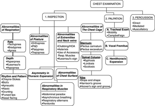
The patient usually remains seated, with the physician moving from front to back and sides. Initial assessment includes an evaluation of effort, rate, and depth of respiration. It also should identify any wheeze, or grunt, or noisy sound that might be audible without the need of a stethoscope. Sequential inspection of the anterior, posterior, and lateral chest should then be carried out. Finally, palpation, percussion, and auscultation complete the exam. The examination should be thorough and go from the chest surface toward the inner structures (Fig. 13-1).
A. Chest Inspection
3 What kind of information can be gathered through inspection?
In addition to gross abnormalities of body habitus (such as the Pickwickian type of obesity-hypoventilation or sleep apnea), an astute examiner also should look for seven important areas of anomaly:
4 What are the most common abnormalities of respiration?
Mostly the ones related to the vital signs of the respiratory pump, such as pattern of breathing and its three main components: (1) rate, (2) depth, and (3) rhythm. Each of them may be abnormal, and all may be evaluated by simply observing the patient during the interview. In addition, grunting, nasal flaring, and pursed-lip respiration also can be detected through bedside observation (see later).
5 What are the abnormalities of posture?
These, too, can be detected by simple observation, even before the patient undresses. They are mostly related to compensatory postures, designed to improve the efficiency of the respiratory “pump.” Patients with chronic obstructive pulmonary disease (COPD), for example, usually sit up and lean forward, so that they can better tense the accessory respiratory muscles and improve their contractility. Patients may lean so much forward to eventually have to prop themselves up by their resting elbows against the thighs. With time, the protracted pressure on the thighs leads to hyperpigmented calluses immediately above the knees (Dahl’s sign).
6 What are the main abnormalities in the use of respiratory muscles?
In addition to recruitment of accessory muscles of respiration, the main abnormality is an asynchronous contraction of the diaphragm and intercostals (paradoxical respiration), an important sign of impending respiratory failure.
7 What about asymmetry in thoracic expansion?
This, too, can be detected by simple inspection, even though it is usually best evaluated by palpation.
8 What abnormalities of the chest cage can be detected by inspection?
Abnormalities of the spine, ribs, and sternum. All may adversely affect lung mechanics and thus lead to compensatory postures.
9 And what about abnormalities of the chest surface?
They are pallor or cyanosis, hyperpigmentation or hypopigmentation, expiratory bulges, collateral circulations, dermatomic lesions, and chest wall fistulas. All are detectable by astute observation.
10 What about assessment of extremities and neck veins?
Although often carried out separately, examination of neck veins and extremities is an important aspect of respiratory evaluation. Finding clubbing, for example, or asterixis may provide valuable information toward the recognition of an underlying respiratory disease. The same can be said for the detection of either nicotine stains (usually on fingers) or a smoker’s face (deep and premature wrinkles coupled with skin coarsening), both indications of unrepentant smoking even before a social history is obtained. Finally, distended neck veins in patients with either cor pulmonale or pulmonary embolism also can be a very important diagnostic sign.
(1) Abnormalities of Respiration
Abnormalities in the Rate of Inspiration
11 What are the main abnormalities in respiratory rate?
The major ones are an increase and a decrease. The normal respiratory rate in an adult should be around 20 ± 5 breaths a minute. Hence, tachypnea indicates a rate faster than 25/minute, whereas bradypnea usually indicates a rate slower than 8/minute, as observed in narcotic-induced respiratory depression.
12 Can tachypnea be considered normal?
Probably not. Still, a respiratory rate >20 breaths/minute is often seen in elderly nursing home residents with chronic medical conditions but no active disease.
13 What is the clinical significance of true tachypnea?
It usually indicates moderate to severe cardiorespiratory disease, requiring a compensatory increase in the work of breathing. In hospitalized patients, it carries a bad prognosis. In fact, on internal medicine wards, it predicts cardiorespiratory arrest. It also argues in favor of pneumonia, both in the inpatient and outpatient settings. In hospitalized pneumonia patients, it can even predict death, thus providing a far more accurate prognostic indicator than either tachycardia or abnormal blood pressure. It also predicts failure to wean from mechanical ventilation.
14 Can absence of tachypnea be helpful?
Yes, because tachypnea is so common in chest diseases that its presence adds relatively little, whereas its absence challenges a cardiac or respiratory diagnosis. For example, tachypnea is so frequent in pulmonary emboli (92% of patients) that a normal respiratory rate argues strongly against the diagnosis. The same can be said for tamponade, where tachypnea is a must. Conversely, in an acute abdomen, tachypnea directs attention to a supradiaphragmatic rather than subdiaphragmatic process.
15 Does tachypnea predict hypoxemia?
Not necessarily. In fact, some patients might actually be hypoxic as a result of hypoventilation (and therefore of bradypnea). Others might use instead tachypnea to fully compensate for hypoxemia. Hence, the need for monitoring not only the respiratory rate, but also oxygen saturation.
16 What is the clinical significance of bradypnea?
It should prompt consideration of hypothyroidism, but it also may suggest a central nervous system disease, or, as indicated before, the use of narcotics and sedatives.
17 What is apnea?
It is the absence of respiration for at least 20 seconds while awake or 30 seconds while asleep. It is often seen in patients with either neuromuscular dysfunction (central apnea) or airway obstruction induced by rapid eye movement (REM) sleep (obstructive sleep apnea). Note that apnea also remains the final event of all respiratory failures, whether due to pulmonary or neuromuscular disease.
Abnormalities in the Depth of Respiration
18 What are the main abnormalities in the depth of respiration?
Hyperpnea and hypopnea (Fig. 13-2).

Figure 13-2 Patterns of respiration. The horizontal axis indicates the relative rates of these patterns. The vertical swings of the lines indicate the relative depth of inspiration.
(Adapted from Seidel HM, Ball JW, Dains JE, Benedict GW: Mosby’s Guide to Physical Examination, 3rd ed. St. Louis, Mosby, 1995.)
19 What is hyperpnea?
It is an increase in tidal volume, usually accompanied (but not necessarily) by an increase in rate. In other words, hyperpnea is a rapid and deep respiration. The classic form was first described by Kussmaul in patients with diabetic ketoacidosis, who attempt to compensate by hyperventilating. This compensation also can be observed in any of the other anion gap metabolic acidoses, which can be recalled with the mnemonic make up a list: methanol poisoning, aspirin intoxication, ketoacidosis, ethylene glycol ingestion, uremia, paraldehyde administration, and lactic acidosis.
20 Is there a difference between hyperpnea and the hyperventilation of cardiorespiratory disease?
Yes. In cardiorespiratory disease, vital capacity is typically compromised, and thus breaths are shallow, with the increase in ventilation due primarily to a faster rate. In contrast, true hyperpnea relies more on increased tidal volume. Since this does not rebreathe dead space as much, it is a better CO2 controller. Hence, hyperpnea rather than tachypnea is the compensation of choice in metabolic acidosis.
21 Who was Kussmaul?
Adolf Kussmaul (1822–1902) was a graduate of Heidelberg and Würzburg (where he studied under Virchow) and a part-time German Army surgeon. He was the first to describe periarteritis nodosa and progressive bulbar paralysis. He also was the first to attempt gastroesophagoscopy, pleural tapping, and peritoneal lavage. His name is linked to the respiration of patients with metabolic acidosis, the clinical description of pericarditis, pulsus paradoxus, aphasia, and, of course, Kussmaul’s sign, the inspiratory increase in jugular venous pressure (and distention) seen in patients with obstruction to right-sided venous return (see Chapter 10, questions 115–118). A meticulous and precise man famous for complaining that none of his colleagues could write good German, Kussmaul contributed satirical poems to a weekly magazine under the pseudonym of Gottlieb Biedermeier, an imaginary and unsophisticated poet who eventually came to symbolize the values and tastes of the early 19th-century German bourgeois: reliable, hard-working, but boringly unimaginative (like in the Biedermeier style of furniture. “Bieder” is German for “everyday, plain” while “Meier,” or Meyer, is a common German last name).
22 What is hypopnea?
It is shallow respiration, usually indicative of impending respiratory failure or obesity-hypoventilation (Pickwickian syndrome). In this regard, hypopnea is often associated with periods of apnea (see question 17).
Abnormalities in Rhythm and Pattern of Respiration
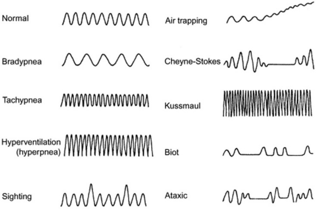
23 What are the main abnormalities in respiratory rhythm?
They are many, and usually the result of disruption in the neurogenic control of the respiratory pump. Hence, they are often seen in comatose patients. Thus, they are valuable to recognize because they may help localizing the site of the neurologic lesion (see Fig. 13-2). Moving downward in a rostrocaudal fashion, from the uppermost to the lowermost neurologic center, the most common abnormalities of respiratory rhythm are (1) Cheyne-Stokes respiration, (2) Biot’s respiration, (3) apneustic breathing, (4) central hyperventilation, and (5) ataxic (agonal) respiration.
24 What is Cheyne-Stokes respiration?
It is a form of periodic breathing (i.e., a regularly irregular pattern consisting of a series of cycles). Each “cycle” has a constant respiratory rate but variable depth, insofar as it progressively increases in amplitude (crescendo), eventually culminating in a peak followed by a decrescendo period. This fades into complete apnea, from which another cycle restarts. The crescendo-decrescendo phase lasts approximately 30 seconds, whereas the apneic period is usually just a little shorter.
25 What is the physiologic repercussion of Cheyne-Stokes?
Mostly a swing in cerebral blood flow, caused by alternating hyperpnea (higher cerebral flow) and hypopnea (lower cerebral flow). These are probably responsible for some of the mental status changes described in these patients, such as alertness, agitation, and increased muscle tone during hyperpnea, followed by sleepiness, motionlessness, and decreased tone during apnea.
26 What is the clinical significance of Cheyne-Stokes?
It may be encountered in normal people as a result of aging or simply sleep.
It also can be seen in normal individuals who recently moved to high altitudes, where environmental hypoxia leads to a heightened CO2 response of the respiratory centers.
The classic association, however, is with congestive heart failure, where Cheyne-Stokes is found in as many as one third of cases, reflecting worse function and prognosis. The reduced cardiac output of these patients leads to a lag between alveolar CO2 and the arterial CO2 delivered to the medulla. Hence, low alveolar CO2 is reflected much later in the blood that bathes the medulla. This asynchrony between alveoli and medulla, coupled to higher sensitivity of the respiratory centers to CO2, eventually leads to the hyperpnea-hypopnea-apnea cycle.
Cheyne-Stokes also can be seen in various neurologic disorders (such as meningitis, bilateral or unilateral cerebral infarctions/hemorrhage, and traumatic brain stem or supratentorial damage) in which the underlying mechanism is heightened sensitivity of the respiratory centers to carbon dioxide stimulations. As a result, any increase in CO2 blood levels leads to excessive hyperventilation, until the CO2 bottoms out and respiration ceases completely. Eventually, the apnea-induced increase in CO2 leads to another phase of hyperpnea, and the cycle starts anew.
27 What are the therapeutic implications of Cheyne-Stokes?
Given its major swings in ventilation (and O2 blood levels), patients with Cheyne-Stokes respiration may require administration of supplemental oxygen and, overall, have a worse prognosis.
28 Who were Cheyne and Stokes?
John Cheyne (1777–1836) was a Scot and himself the son of a surgeon, often helping his father to care for patients by bleeding and dressing them. After graduating at age 18 from the University of Edinburgh, he served in the army for 4 years. During this time, he took part in the battle of Vinegar Hill, which broke Irish resistance to British rule. In 1809, he went to Dublin, where he was eventually appointed Physician-in-General for Ireland, becoming the founder of modern Irish medicine.
William Stokes (1804–1878) was instead a bona-fide Irishman and the son of the anatomy professor who had succeeded John Cheyne at the College of Surgeons School in Ireland. Although lacking in formal education (his father wanted to protect him from a society that did not abide by the scriptures), Stokes eventually went to Edinburgh, where he received his doctor of medicine in 1825. In Scotland, he learned of Laënnec and his recent invention, the “cylinder.” He soon became so enamored of this little tool that he even wrote an introductory book about it, the first of its kind in the English language. In fact, Stokes was such a vocal advocate for the use of stethoscopy that he provoked quite a few reactions (and even some sarcasm) among his colleagues. Still, he was a well-liked physician, who worked among the poor during the Dublin typhus epidemic in 1826 (he even contracted the disease but survived) and then again during the subsequent cholera epidemic. His name is linked not only to the eponymous pattern of respiration but also to Stokes-Adams syncope, which the Irish surgeon Robert Adams had described in 1827 and which Stokes included in his 1854 book, Diseases of the Heart and Aorta. Of course, the Italian Morgagni had preceded them both by describing the condition almost 100 years before (see Chapter 11, question 14).
29 What other abnormalities in rhythm are worthy of recognition? What is their significance?
Biot’s respiration is a variant of Cheyne-Stokes, insofar as it is a succession of hyperpneas/hyperventilations and apneas, but without the typical crescendo-decrescendo pattern, the abrupt beginning, and the regularity. It is also less common than Cheyne-Stokes. Biot’s is usually observed in patients with either meningitis or medullary compression. Hence, it carries a worse prognosis, usually resulting in complete apnea and cardiac arrest.
Apneustic breathing is a peculiar pattern of respiration, characterized by a deep inspiratory phase followed by a breath-holding period and a rapid exhalation. It is typical of brain stem (pons) lesions.
Central hyperventilation is often encountered in patients with midbrain/upper pontine lesions. It is an ongoing pattern of hyperpnea and tachypnea (i.e., deep and rapid respirations), which tends to be different from Kussmaul’s respiration, insofar as it is not as fast, but usually deeper.
Ataxic ventilation: From the Greek a- (lack of) and taxis (order), this is a totally anarchic respiratory rhythm—a sort of fibrillation of the respiratory centers, with back-and-forth shifts from hyper- to hypo-ventilation, and from hyperpnea to hypopnea, all intermingled with periods of apnea. It is seen in patients with damage to the medulla, typically preceding death. Hence the term, agonal respiration.
30 What is a grunting respiration?
It is another abnormal respiratory pattern. A typical grunting respiration is the râle de la mort, a pre-terminal gurgling-and-grunting sound produced by patients too ill to clear secretions. The expression translates as “death rattle” (râle is rattle in French), and in the olden days, it used to be a sign of severe pneumonia with impending respiratory muscle fatigue and death. The râle de la mort also is the main cause of Laënnec’s botched nomenclature, insofar as the inventor of the stethoscope became so sensitive to the emotional overtones of the term râle to eventually prefer at the bedside its Latin equivalent rhonchus. This triggered a tremendous (and ongoing) confusion in lung sound terminology.
31 What is the clinical significance of a grunting respiration?
Very much the same as in Laënnec’s days. It can still be heard in adults with respiratory muscle fatigue (and impending arrest), but nowadays it is much more frequent in children, where it usually presents as a short and low-pitched noise produced by forced expiration against a closed glottis. The “grunt” is due to the sudden opening of the glottis and the loud rush of air from the larynx. Its physiology (and significance) is akin to pursed-lip respiration (see below, questions 32 and 33), insofar as it leads to an increase in expiratory airway pressure, which then acts as a mechanical splint against alveolar collapse, increasing tidal volume and oxygenation while decreasing respiratory rate and CO2. An increased intra-alveolar pressure also has a positive effect against transudation of fluid in patients with pulmonary edema, and thus it is often observed during acute episodes of left ventricular failure.
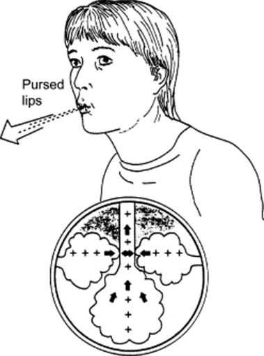
32 What is pursed-lip respiration?
Another respiratory pattern, typically seen in obstructive lung disease—usually emphysema (Fig. 13-3). Given the alveolar hyperinflation (and reduced lung elasticity) of COPD, patients are at risk for expiratory airway closure and air-trapping. Hence, they resort to pursed lip exhalation, as if they were inflating a balloon. This increases intra-airway pressure, thus inducing auto-PEEP (positive end-expiratory pressure). It is often accompanied by an expiratory wheeze or grunt.

Figure 13-3 Demonstration of pursed-lip breathing in patients with COPD and its effects. The weakened bronchial airways are kept open by the effects of positive pressure created by pursed lips during expiration.
(From Hillegass EA, Sadowsky HS [eds]: Essentials of Cardiopulmonary Physical Therapy. Philadelphia, WB Saunders, 1994.)
33 What is the physiologic impact of pursed-lip respiration?
It increases arterial O2 while decreasing CO2. It does so by behaving as an E-PAP in a Bi-PAP machine (i.e., by providing an expiratory positive airway pressure). It also slows respiratory rate (by as much as 40%), improves tidal volume, and decreases dyspnea. The mechanism for the latter is unclear, although it probably involves an effect on interstitial J-receptors as well as an overall reduction in the work of breathing.
34 What is nasal flaring?
It is the outward inspiratory motion of the nostrils—a valuable sign of respiratory distress. Often associated with the use of accessory muscles.
36 What is orthopnea?
It is a dyspnea that is aggravated by lying flat. Conversely, it is relieved by sitting upright. It comes from the Greek orthos (upright) and pneo (breathing)—(i.e., upright respiration).
37 What is the clinical and physiologic significance of orthopnea?
Usually indicates congestive heart failure, where it can help identify patients with low ejection fraction. Conventional wisdom has traditionally interpreted orthopnea as a “poor man’s” phlebotomy, pooling blood in the dependent areas of the body and thereby decreasing venous return and ventricular preload. Yet, in patients with congestive heart failure, orthopnea and pulmonary capillary wedge pressure correlate very little. Still, the need for orthopnea is often relieved whenever left ventricular failure degenerates into bi-ventricular failure, suggesting that failure of the right ventricle may indeed provide a useful “unloading” to left ventricular filling and pulmonary congestion.
38 Can orthopnea also occur in patients with lung disease?
Yes. Although more common in heart disease (in as many as 95% of cases), orthopnea also may be observed in lung disease, since sitting upright improves both vital capacity and lung compliance. Hence, orthopnea can occur in many pulmonary conditions, such as pneumonia, bilateral diaphragmatic paralysis, and pleural effusion. It also can occur in bilateral apical lung diseases, usually bullous. Whenever these patients sit up, they increase perfusion to the lower lung fields (as a result of gravity). Since these areas also are the best ventilated (because of bilateral apical disease), orthopnea improves ventilation/perfusion matching and gas exchange and thus relieves dyspnea.
39 And what about patients with COPD?
In COPD (which often presents with apical bullae), an upright position improves not only gas exchange but also lung mechanics (because of the increased stretch of accessory respiratory muscles). Hence, COPD patients often tend to prop themselves up, so that they can better use their respiratory muscles. They do so by clasping the side of the bed or pushing with the elbows over the thighs (see question 5, Dahl’s sign).
40 What about asthma?
Orthopnea also is an important sign of asthma, especially severe asthma. In fact, when present at the time of initial emergency evaluation, it is a good predictor of poor outcome. Patients who cannot lie flat have a worse pulmonary function and a greater need for admission. The same is true for diaphoresis. Both findings were reported by Brenner in acute asthmatics and represent the scientific validation of the time-honored dictum that patients who “do not look good” (because they are sweaty and in an obligatory upright position) usually do poorly.
41 So, is orthopnea a cardiac or a pulmonary sign?
It is both. Because of all the above reasons, orthopnea should not be interpreted as a sign of a cardiac rather than respiratory etiology. The contrary is actually the case. Yet, absence of orthopnea in pulmonary patients does argue against concomitant presence of left ventricular failure.
42 Can orthopnea be encountered in patients with neither cardiac nor pulmonary disease?
Yes. For instance, it can be seen in patients with massive ascites, obesity, or bilateral phrenic nerve paralysis. In both cases, an upright position helps relieve the intra-abdominal pressure on the lungs.
43 What is PND?
It is a very common and dramatic presentation, characterized by a nocturnal spell (paroxysm) of acute dyspnea (air hunger). After 1–2 hours of sleep, the patient suddenly awakens, sits upright, lowers the legs down the side of the bed, opens the window to catch some fresh air, and after a few minutes feels better enough to go back to sleep.
44 What is the mechanism of relief in PND?
It is the upright posture, of course, and not the fresh air (although cold air blown into the face of patients has been shown to give a refreshing feeling in both cardiac and pulmonary diseases). The peripheral blood-pooling caused by sitting upright effectively decreases venous return, thereby reducing pulmonary capillary pressure and lung congestion. PND is, therefore, a frequent sign of left ventricular failure.
45 Can PND be seen in pulmonary patients, too?
Yes. Like orthopnea, it can be seen in bullous and bilateral apical disease. Both basilar perfusion and lung mechanics are improved when the patient sits upright and leans forward.
46 What is platypnea?
From the Greek platus (flat) and pneo (respiration), platypnea is an obligatory supine respiration. In other words, patients with platypnea breathe better when they lie flat. Hence, it is the opposite of orthopnea: it is a dyspnea in the erect position, promptly relieved by recumbency. Platypnea is often associated with orthodeoxia, which is a hemoglobin oxygen desaturation upon sitting upright.
47 What is the clinical significance of platypnea?
Unlike orthopnea, platypnea is usually due to a right-to-left shunt, which can be either intracardiac or intrapulmonary. In intrapulmonary shunts, platypnea requires a bibasilar process (and not biapical, like orthopnea). In this case, an upright posture increases perfusion to the lower lobes, worsens ventilation/perfusion (V/Q) matching, and leads to oxygen desaturation and dyspnea. Conversely, a supine position improves V/Q matching and relieves dyspnea. Given its physiology, platypnea occurs in:
Multiple recurrent pulmonary emboli (which, because of gravity, tend to involve primarily the bases)
Pleural effusion (which can cause bibasilar atelectasis) or bibasilar pneumonia
Cirrhosis (which often has arteriovenous shunting at the lung bases)
Intrapulmonary right-to-left shunt, such as an arteriovenous malformation, often basilar in location
Intracardiac right-to-left shunt, usually due to an atrial septal defect. This produces platypnea only when associated with an increase in pulmonary resistances, as in cases of pleurocardial/pericardial effusion or status post lobectomy/pneumonectomy. An upright position will reduce the shunt by redirecting blood toward the atrial septum and by possibly increasing pressure over the right atrium. A supine posture will have instead the opposite effect.
48 What is trepopnea?
From the Greek trepo (twisted) and pneo (breathing), this is a “twisted respiration” characterized by the patient’s inability to lie supine (or prone), and instead by preference for the lateral decubitus position. Trepopnea is often referred to as “down with the good lung,” meaning that in cases of unilateral lung disease, the patient can breathe better when placed on a side, typically with the good lung down.
49 What is the physiology behind trepopnea?
It is an increased perfusion to the dependent lung, which happens to be the good one, thus causing better (V/Q) matching, better oxygenation, and more comfortable respiration.
50 What are some of the disease processes associated with trepopnea?
The classic one is a unilateral lung collapse, from an endobronchial obstructing lesion or a massive effusion. In both situations, the patient feels better (and has improved oxygenation) whenever the good lung is “dependent.” A similar mechanism also can explain the preferential right lateral decubitus position of patients with congestive heart failure from dilated cardiomyopathy (since this relieves the pressure applied on the left lung by the enlarged heart), or the preferential lateral decubitus position of patients with a mediastinal or endobronchial tumor that compresses the airways only in a particular position.
51 Are there any contraindications to lying with the “good” lung down?
Any unilateral lung disease due to “spillable” material, like pneumonia (intra-alveolar pus) or hemorrhage (intra-alveolar blood). In this case, intrabronchial spread into the dependent good lung would only make things worse. Hence, patients with “spillable” unilateral disease should always lie with the bad lung down, since protecting the good lung is more important than improving oxygenation. Finally, lying with the good lung down is physiologically detrimental in small children with unilateral lung disease.
(3) Abnormalities in the Use of Respiratory Muscles
52 What are these?
These are abnormalities in the respiratory muscle “pump,” characterized by weakness and fatigue, eventually leading to respiratory failure. They involve primarily the diaphragm but also can affect the intercostals. They are (1) paradoxical respiration (abdominal paradox) and (2) respiratory alternans.
53 What is abdominal paradox?
It is a sign of diaphragmatic fatigue. During normal respiration, the abdominal and thoracic wall are synchronized, both expanding in inspiration and contracting in exhalation (even though chest expansion is usually greater in the upright position, whereas abdominal expansion is usually greater in the supine position). In some conditions, however, the chest and abdomen become asynchronous, with the chest expanding in inspiration while the abdomen is instead being pulled in and vice versa. This “rocking motion” (labeled abdominal paradox, paradoxical respiratory breathing, or respiratory paradox) usually indicates bilateral diaphragmatic weakness or paralysis, causing the diaphragm to behave as a passive membrane, sucked up into the chest during inspiration and pushed down into the abdomen during exhalation.
54 What is the best way to detect abdominal paradox?
Bimanual palpation. Lay one hand over the patient’s chest and one over the abdomen—then look for rocking motion. Note, however, that “paradox” also can be detected by simple inspection.
55 Are patients with abdominal paradox orthopneic?
Yes. Since in a supine position the abdominal content applies greater pressure on the diaphragm, patients with abdominal paradox try to compensate by assuming an upright posture.
56 How clinically valuable is this maneuver in predicting respiratory failure?
Quite valuable. Abdominal paradox has high sensitivity (95%) and good specificity (71%). In fact, in impending respiratory failure, it usually precedes deterioration of arterial blood gases.
57 What is asynchronous breathing?
It is a special form of abdominal paradox, seen in patients with chronic obstructive lung disease. In this case, the inward movement of the abdominal wall in early exhalation is almost immediately followed by an outward movement. This particular form of expiratory “rocking” reflects worse pulmonary function and prognosis, predicting respiratory failure, requirement for mechanical ventilation, and death.
58 What is respiratory alternans?
Another sign of respiratory muscle weakness and impending failure. It may occur in place of, or in combination with, abdominal paradox. Patients with respiratory alternans exhibit alternate use of either the diaphragm or intercostals, with the chest and abdomen first “rocking” one way, and then the other way. Occasionally, patients may cycle from respiratory alternans to abdominal paradox and vice versa.
59 How is respiration in patients with peritonitis?
Limited. In peritonitis, the abdominal wall can become quite still during respiration. This limitation in movement is diffuse in generalized peritonitis and localized in focal involvement. In diverticulitis, for example, the motionless area is the left lower quadrant; in appendicitis it is the right lower quadrant.
(4) Asymmetry in Thoracic Expansion
60 Can inspection identify an asymmetry in thoracic expansion?
Yes. Although not as effective as palpation, inspection can identify asymmetries of expansion between the two hemithoraces. These may occur in many lung conditions (including atelectasis, pneumonia, and pleural effusion), although it is only in the most severe cases (such as a large pneumothorax, a complete lung collapse, or a massive effusion) that the degree of change in volume of one hemithorax becomes large enough to be detectable by inspection. Protracted lung collapse may even lead to a deviation in spinal curvature, causing a concavity toward the side of disease.
61 What is the best way to identify a thoracic asymmetry by inspection alone?
Deep inspiration, since these lags are typically undetectable during quiet respiration. Hence, ask the patient to take a full deep breath and then look for a local lagging in chest expansion.
63 What are the main chest cage abnormalities?
It depends on whether they involve the spine, sternum, or ribs. Often, more than one component is affected.
If the spine is affected, the abnormalities may be on the sagittal or frontal plane.
If the sternum is involved, the two most common abnormalities are pigeon chest and barrel chest. Both may alter lung mechanics severely enough to reduce lung function.
If ribs are involved, the most common abnormalities concern costal slope and shape.
65 What abnormalities may be seen on the sagittal plane?
These are an increase in spinal convexity (lordosis) or concavity (kyphosis)—often coexisting.

Stay updated, free articles. Join our Telegram channel

Full access? Get Clinical Tree

