Anatomy of the Brain
The brain contains millions of neurons arranged in a vast array of synaptic connections that provide seemingly unfathomable circuitry. Through that circuitry, the brain integrates and processes sensory information and provides motor output.
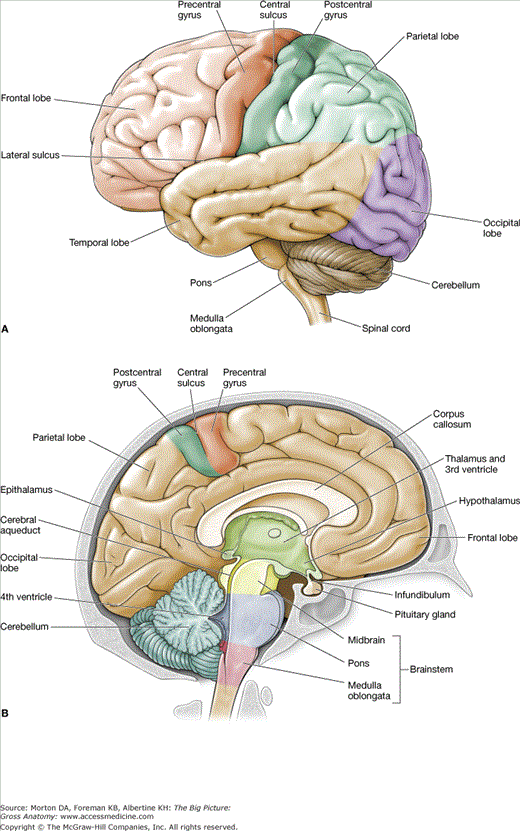
The brain is divided into the cerebrum, diencephalon, brainstem, and cerebellum (Figure 16-1A and B).
The cerebrum is the organ of thought and serves as the control site of the nervous system, enabling us to possess the qualities associated with consciousness such as perception, communication, understanding, and memory (Figure 16-1A). The cerebral hemispheres consist of elevations (gyri) and valleys (sulci), with a longitudinal cerebral fissure separating the hemispheres. Each cerebral hemisphere is divided into lobes, which correspond roughly to the overlying bones of the skull.
- Frontal lobe. The frontal lobe is located in the anterior cranial fossa. The central sulcus divides the frontal lobe from the parietal lobe in a coronal plane. The gyrus anterior to the central sulcus is called the precentral sulcus and serves as the primary motor area of the brain. The remainder of the frontal lobe is used in modifying motor actions.
- Parietal lobe. The parietal lobe interprets sensations from the body. The gyrus posterior to the central sulcus, the postcentral sulcus, is the primary area for receipt of these sensations.
- Occipital lobe. The occipital lobe is located superior to the tentorium cerebelli, in the posterior cranial fossa, and is primarily concerned with vision.
- Temporal lobe. The temporal lobe is located in the middle cranial fossa and is primarily concerned with hearing.
The diencephalon consists of the thalamus, hypothalamus, epithalamus, and subthalamus, and is situated between the cerebrum and the brainstem (Figure 16-1B). The diencephalon serves as the main processing center for information destined to reach the cerebral cortex from the ascending pathways.
The brainstem consists of the midbrain, pons, and medulla oblongata (Figure 16-1B).
- Midbrain. The midbrain (mesencephalon) contains the nuclei for the oculomotor nerve and the trochlear nerve, cranial nerves (CNN) III and IV, respectively. The cerebral aqueduct is a portion of the ventricular system and courses through the center of the midbrain to connect the third and fourth ventricles.
- Pons. The pons is situated against the clivus and the dorsum sellae and contains the nuclei for the trigeminal, abducens, facial, and vestibulocochlear nerves (CNN V, VI, VII, and VIII, respectively).
- Medulla oblongata. The medulla oblongata, commonly called the medulla, is located at the level of the foramen magnum. It serves as the major autonomic reflex center that relays visceral motor control to the heart, blood vessels, respiratory system, and gastrointestinal tract. It possesses the nuclei for the glossopharyngeal, vagal, accessory, and hypoglossal nerves (CNN IX, X, XI, and XII, respectively).
The cerebellum lies in the posterior cranial fossa and assists in the coordination of skeletal muscle contraction (Figure 16-1A and B). It functions at a subconscious level and provides skeletal muscles with precise timing and appropriate patterns of contraction needed for smooth, coordinated movements.
Ventricular System of the Brain
Stay updated, free articles. Join our Telegram channel

Full access? Get Clinical Tree


