Pericardium
The pericardium is a sac that encloses the heart, akin to the pleura that encloses the lungs. The pericardium and the heart are located in the middle of the thorax, between T4 and T8 vertebrae. The pericardium has parietal and visceral layers.
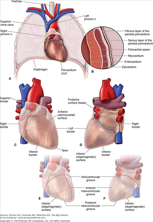
The parietal pericardium is composed of an external fibrous layer (fibrous pericardium) and an internal serous layer (serous pericardium) (Figure 4-1A and B).
- Fibrous pericardium. A strong, dense collagenous tissue that blends with the tunica externa of the great vessels and the central tendon of the diaphragm.
- Serous pericardium. Lines the inner surface of the fibrous pericardium.
- Pericardial space. Lies between the serous layer of the parietal pericardium and the visceral pericardium.
- Visceral pericardium (epicardium). A serous layer that intimately follows the contours of the heart surface. At the root of the heart, the visceral pericardium is contiguous with the serous pericardium, analogous to the visceral and parietal pleura of the hilum of each lung.
The pericardial sinuses are subdivisions of the pericardial sac and consist of the following spaces:
- Transverse sinus. Lies posterior to the ascending aorta and the pulmonary trunk and anterosuperior to the left atrium and the pulmonary veins.
- Oblique sinus. Lies posterior to the heart and is surrounded by the reflection of the serous pericardium around the right and left pulmonary veins and the inferior vena cava.
The pericardium receives its blood supply from the pericardiacophrenic vessels and branches from the bronchial and esophageal vessels. It is innervated by visceral sensory fibers from the phrenic and vagus nerves and the sympathetic trunks.
Overview of the Heart
The heart has the following three layers (Figure 4-1B):
- Epicardium. The outer layer, also known as the visceral pericardium.
- Myocardium. The middle layer, consisting of cardiac muscle responsible for contraction of the heart.
- Endocardium. The inner layer, consisting of endothelial cells that line the lumen of the four chambers.
The heart, including its left and right sides, surfaces, borders, and sulci (Figure 4-1C to F), art described in a variety of ways.
The expression “right side of the heart” refers to the right atrium and the right ventricle, which collect systemic deoxygenated blood and pump it into the lungs. By comparison, the “left side of the heart” refers to the left atrium and the left ventricle, which collect oxygenated blood from the pulmonary circulation and pump it into the body.
The shape of the heart is roughly that of a cone with a flat base and a pointed apex. The surfaces of the heart consist of the following:
- Base. Is flat, faces posteriorly, and is formed mainly by the pulmonary veins of the left atrium. The base faces posteriorly toward the T6–T9 vertebral bodies, intervened by the pericardium, the esophagus, and the aorta.
- Sternocostal surface. Is part of the heart’s cone shape and is named for its location deep to the sternum and costal cartilages of the five uppermost ribs.
- Diaphragmatic surface. Is also part of the heart’s cone shape and is named for the heart surface directly superior to the central tendon of the diaphragm. Note that the heart, as it lies within the thorax, rests upon the diaphragmatic surface but not on its base.
The apex is formed by the tip of the left ventricle and points anteriorly, inferiorly, and to the left. The apex lies deep to the left fifth intercostal space medial to the midclavicular line. In an adult, the apex of the heart is approximately one hand’s breadth from the median plane, near the midclavicular (nipple) line. This location is useful clinically because auscultation of the mitral valve is best heard over the apex.
- The superior border is formed by the right and left auricles. The ascending aorta and pulmonary trunk emerge from this border, and the superior vena cava enters the right atrium.
- The right ventricle forms the inferior border.
- The left border is formed by the left ventricle and auricle of the left atrium.
- The right border is formed by the superior vena cava, right atrium, and inferior vena cava.
Sulci (grooves) mark the outer surface of the heart at the sites where one chamber meets another. The cardiac sulci are as follows:
- Sulcus terminalis. A groove on the external surface of the right atrium. This sulcus marks the junction of the primitive sinus venosus with the atrium in the embryo, and corresponds to a ridge on the internal surface of the right atrium, the crista terminalis.
- Atrioventricular (AV) groove (coronary sulcus). An external demarcation of the atria from the ventricles. The AV groove contains the right coronary, the left coronary, and the circumflex coronary arteries and the coronary sinus.
- Anterior interventricular groove. Is demarcates the left ventricle from the right ventricle on the anterior surface of the heart and contains the left anterior descending artery and the great cardiac vein.
- Posterior interventricular groove. Is demarcates the left ventricle from the right ventricle on the posterior surface of the heart and contains the posterior interventricular artery and the middle cardiac vein.
Coronary Circulation
Although blood fills the chambers of the heart, the myocardium is so thick that it requires its own artery–capillary–vein system, called the “coronary circulation,” to deliver and remove blood to and from the myocardium. The vessels that supply oxygenated blood to the myocardium are known as coronary arteries. The vessels that remove the deoxygenated blood from the heart muscle are known as cardiac veins.
The coronary arteries and branches course along the epicardium in the cardiac sulci and interventricular grooves (Figure 4-2). Each coronary artery sends branches to the heart muscle.
Stay updated, free articles. Join our Telegram channel

Full access? Get Clinical Tree


