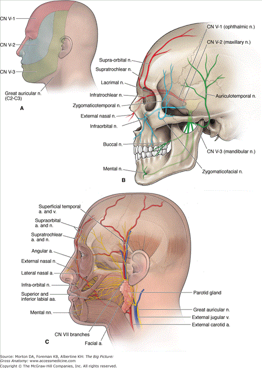
Cutaneous Innervation and Vasculature of the Face
The sensory innervation of the face is provided by the three divisions of the trigeminal nerve [cranial nerve (CN) V], with each division supplying the upper, middle, and lower third of the face. The facial artery and the superficial temporal artery provide vascular supply.
The skin of the face and scalp is innervated by the cutaneous branches of the three divisions of CN V and by some nerves from the cervical plexus (Figure 20-1A and B).
- CN V-1 (ophthalmic nerve). Provides cutaneous innervation to the anterior region of the scalp via the supraorbital nerve and the supratrochlear nerve, the skin of the upper eyelid via the lacrimal nerve, and the bridge of the nose via the external nasal nerve and the infratrochlear nerve.
- CN V-2 (maxillary nerve). Provides cutaneous innervation to the skin along the zygomatic arch via the zygomaticofacial and the zygomaticotemporal nerves and the skin of the maxillary region, lower eyelid, and upper lip via the branches of the infraorbital nerve.
- CN V-3 (mandibular nerve). Provides cutaneous innervation to the skin of the lateral aspect of the scalp and the lateral part of the face, anterior to the external acoustic meatus, via the auriculotemporal nerve, the skin covering the mandible via the buccal nerve, and the skin of the lower lip via the mental nerve.
- Cervical plexus. The great auricular nerve (C2–C3) innervates the skin over the angle of the mandible just in front of the ear.
 Trigeminal neuralgia (tic douloureux) is a condition marked by paroxysmal pain along the course of CN V. Sectioning the sensory root of CN V at the trigeminal ganglion may alleviate the pain.
The face has a very rich blood supply provided primarily from the following vessels (Figure 20-1C):
- Facial artery. Branches off the external carotid artery and courses deep to the submandibular gland; winds around the inferior border of the mandible, anterior to the masseter muscle, to enter the face. As it ascends in the face, the facial artery supplies most of the face by way of the inferior labial, superior labial, lateral nasal, and angular arteries.
- Supraorbital and supratrochlear arteries. Terminal branches of the ophthalmic artery, a branch of the internal carotid artery, which supply the anterior portion of the scalp.
- Superficial temporal artery. A terminal branch of the external carotid artery provides arterial supply to the lateral surface of the face and scalp.
- Facial vein. Formed by the union of the supraorbital and supratrochlear veins. The facial vein descends in the face and receives tributaries corresponding to the branches of the facial artery. The facial vein drains into the internal jugular vein.
 In the skull, the facial vein connects with the cavernous sinus via the superior ophthalmic vein. Unlike other systemic veins, the facial and superior ophthalmic veins lack valves, thus providing a potential pathway for the spread of infection from the face to the cavernous sinus.
Stay updated, free articles. Join our Telegram channel

Full access? Get Clinical Tree


