Female Reproductive System
The female reproductive system consists of the ovaries, uterine tubes, uterus, vagina, and external genitalia. These organs remain underdeveloped for about the first 10 years of life. During adolescence, sexual development occurs and menses first occur (menarche). Cyclic changes occur throughout the reproductive period, with an average cycle length of approximately 28 days. These cycles cease at about the fifth decade of life (menopause), at which time the reproductive organs become atrophic.
The ovaries are the primary female sex organ because they produce eggs (ovum or oocytes) and sex hormones (e.g., estrogen). The ovaries are located within the pelvic cavity.
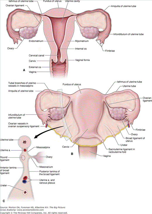
The paired uterine tubes are also called fallopian tubes, or oviducts, and extend from the ovaries to the uterus (Figure 14-1A and B). The luminal diameter of the uterine tubes is very narrow and, in fact, is only as wide as a human hair.
- Infundibulum and fimbriae. The infundibulum is the funnel-shaped, peripheral end of the uterine tube. The infundibulum has fingerlike projections called fimbriae. The fimbriated end of the uterine tube is not covered by peritoneum, which provides open communication between the uterine tube and the peritoneal (pelvic) cavity. In contrast to the male reproductive system, where the tubules are continuous with the testes, the uterine tubes are separate from the ovaries. Oocytes are released (ovulation) into the peritoneal cavity. The beating of the fimbriae may create currents in the peritoneal fluid, which carry oocytes into the uterine tube lumen.
- Ampulla. The ampulla is a region of the uterine tube where fertilization usually occurs.
- Isthmus. The isthmus is the constricted region of the uterine tube where each tube attaches to the superolateral wall of the uterus.
 An ectopic pregnancy occurs when a fertilized egg implants in the uterine tube or peritoneal cavity. The uterine tubes are not continuous with the ovaries, and therefore, there is also a risk that fertilization and implantation will occur outside of the uterine tubes in the peritoneal cavity. Ectopic pregnancies usually result in loss of the fertilized ovum and in hemorrhage, putting the health of the woman at risk.
The uterus, known as the womb, resembles an inverted pear and is located in the pelvic cavity between the rectum and the urinary bladder (Figure 14-1A and B). The uterus is a hollow organ that functions to receive and nourish a fertilized oocyte until birth. Normally, the uterus is flexed anteriorly, where it joins the vagina; however, the uterus may also be retroverted (flexed posteriorly). The pelvic and urogenital diaphragms support the uterus. The uterus consists of the following subdivisions:
Stay updated, free articles. Join our Telegram channel

Full access? Get Clinical Tree


