MOVEMENT THROUGH THE MEMBRANE
Lipid-soluble substances, such as oxygen, carbon dioxide, alcohol, and urea, move across the lipid bilayer by simple diffusion. Other substances that are not lipid soluble, such as most small ions, glucose, amino acids, and proteins, move between the extracellular fluid and the intracellular compartments through pores provided by the integral proteins or through carrier-mediated transport systems. Carrier-mediated transport also originates in the integral proteins. The extracellular fluid consists of the fluid between the cells, called the interstitial fluid, and the blood. The fluid inside the cell is called the intracellular fluid.
Simple Diffusion through the Cell Membrane
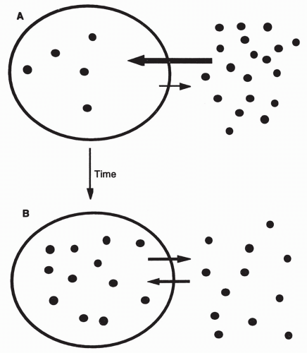
Simple diffusion occurs through random movement of molecules from an area of higher concentration to an area of lower concentration until equilibrium is reached (
Fig. 1-2). This process does not require energy, but can result in the movement of a substance across a membrane. However, the substance cannot accumulate in higher concentration on one side of the membrane compared to the other.
Osmosis
The movement of water across a semipermeable membrane from an area of lower particle concentration to an area of higher concentration is called osmosis. The drive for water to move in one direction or the other is described as the
osmotic pressure. The osmotic pressure of a solution depends on the number of particles or ions present in the water solution and the
hydrostatic pressure (mechanical water force against cell membranes). The more ions are
present in the solution, the lesser is the water concentration and the greater the osmotic pressure (i.e., the pressure for water to diffuse into the solution). Osmosis continues until equilibrium is reached or hydrostatic pressure opposes the flow.
Simple Diffusion through Protein Pores
Small ions, such as hydrogen, sodium, potassium, and calcium, are too electrically charged to diffuse through the lipid membrane of the cell. Instead, they diffuse through the pores provided by the integral proteins. Selectivity of the protein channels is based on the shape and size of the channel and the electrical nature of the ion.
Many protein channels are gated; they can be open or closed to an ion. Whether the gate is open or closed usually depends on the electrical potential across the gate (i.e., the voltage-gated sodium channel), or on binding to the
gate by a ligand that causes it to open or close. An example of ligand gating is when acetylcholine binds to proteins on the neuromuscular junction, thereby opening gates to many small molecules, especially sodium and, to a lesser extent, calcium ions. Diffusion through a gate continues until the concentrations on either side of the membrane are equal or the gate is closed. Several human diseases are related to dysfunction of transmembrane protein channels. Cystic fibrosis is the best-known human disease caused by a defective transmembrane protein that results in abnormal ion movement through a pore.
Mediated Transport
For many substances like glucose and amino acids, simple diffusion is impossible. These molecules are too charged to pass through the lipid portion of the membrane or too large to pass through a pore. Instead, these substances, called substrates, are transported across the membrane with the assistance of a carrier. This type of movement is called mediated transport and may require energy derived from the splitting of adenosine triphosphate (ATP) (see Energy Production section).
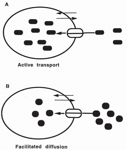
Active transport is mediated transport that requires energy (
Fig. 1-3A). With active transport, energy is used by the cell to maintain a substance at higher concentration on one side of the membrane than the other. Examples of substances moved by active transport include sodium, potassium, calcium, and the amino acids. Each of these substances is actively transported, with the assistance of a carrier, in one direction against a concentration gradient. It then moves down its concentration gradient by simple diffusion in the opposite direction.
Facilitated diffusion is mediated transport that does not require energy, but cannot concentrate a substance (
Fig. 1-3B). Simple diffusion continues at some level in all carrier-mediated systems. Facilitated diffusion is similar to simple diffusion in that no energy is used by the cell to transport a substance; therefore, the substance cannot be transported against its concentration gradient. With facilitated diffusion, a molecule that is limited in its ability to cross the cell membrane on its own is assisted (facilitated) by a carrier to cross the membrane. For example, insulin increases the transport of glucose into most cells.
The Sodium-Potassium Pump
An important example of active transport is the pumping of sodium and potassium across cell membranes. This transport depends on an integral carrier protein known as the sodium-potassium pump. Associated with the pump is an enzyme that splits ATP and provides the energy needed for the pump to function. This enzyme is known as the sodium-potassium ATPase. The sodium-potassium pump transports sodium ions out of the cells to the extracellular region where the concentration is approximately 14 times that inside
the cell. Potassium ions are moved into the cell where the concentration is approximately 35 times greater than outside the cell. This transport causes greater sodium concentration in the extracellular fluid (142 mEq/L) compared to the intracellular fluid (14 mEq/L), and greater potassium concentration in the intracellular fluid (140 mEq/L) compared to the extracellular fluid (4 mEq/L). The sodium-potassium pump carries three sodium molecules out of the cell for every two potassium molecules it carries in.
The Effects of Pumping Sodium and Potassium
Because sodium and potassium are cations (carrying a positive charge), the transport of three sodium ions out of the cell and only two potassium ions into the cell creates an electrical gradient across the cell membrane. It is this electrical
membrane potential that allows nerve and muscle function and action potentials to occur (see
Chapter 8).
The sodium-potassium pump is essential in controlling cell volume. The presence of intracellular proteins and other organic substances that cannot cross the cell membrane increases intracellular osmotic pressure and creates
a tendency for water to diffuse into the cell. This diffusion of water, if unlimited, would cause the cell to swell and eventually burst. However, with the active transport of three sodium ions out of the cell, the osmotic pressure inside the cell is reduced and the diffusion of water into the cell is contained.
Coupled Transport
Many substances are transported coupled to the active transport of sodium. These substances include glucose, amino acids, hydrogen ions, and calcium. The energy required for the transport of these other substances is indirectly supplied through the splitting of ATP by the sodium-potassium ATPase. This type of transport is called secondary active transport.
Coupled transport can be in the same direction as sodium transport (cotransport) or it may occur in the direction opposite to that of sodium transport (countertransport). Both types of transport depend on the diffusion of sodium down its concentration gradient, which in turn depends on the active transport of sodium by the sodium-potassium pump. In cotransport, sodium attaches to a carrier that assists it in moving down its concentration gradient, from outside the cell to inside; the other substance attaches to the same carrier, also on the outside of the cell, and as sodium is moved into the cell, the coupled substance moves with it. In countertransport, the coupled substance binds to the sodium carrier on the inside of the cell membrane with sodium on the outside. Therefore, when sodium is delivered to the inside of the cell, the other substance is delivered to the outside.
Calcium Transport
Calcium can move by simple diffusion through the cell membrane, be transported by coupled transport with sodium, or be moved by primary active transport through a calcium pump. There are two known calcium pumps. One is part of an integral protein present in the cell membrane, which moves calcium out of the cell. The other is an intracellular pump, which pumps calcium out of the cytoplasm into intracellular compartments such as the sarcoplasmic reticulum. This results in sequestering calcium inside the cell. Both pumps keep free intracellular calcium concentration low. The calcium pumps serve as ATPases, which derive energy needed to pump the calcium against its concentration gradient by the splitting of ATP.
Characteristics of Carriers
Active transport and facilitated diffusion require carriers. All carriers are affected by the properties of specificity, saturation, and competition.
Specificity of carriers means that only certain substrates will be transported by any one carrier. The carrier and its specific substrate appear to fit together like a lock and a key.
Saturation of carriers means that at a certain concentration of substrate, all carriers will be filled and transport will level off. Additional substrate will not increase transport across the membrane.
Competition of carriers occurs when there is more than one substrate transported by the same carrier. The multiple substrates compete with each other for the limited number of carrier sites available. Many drugs, naturally occurring and synthetic, compete with endogenous hormones and neurotransmitters for various carrier molecules.
Endocytosis and Exocytosis
When large substances cannot enter the cell by diffusion or mediated transport, endocytosis (engulfment) of the substance by the cell membrane occurs. Pinocytosis is the engulfment of, fluids and small particles by vesicles and is important in the transport of proteins to the cytoplasm. Phagocytosis involves the engulfment and degradation of microorganisms such as bacteria. Both processes require energy. Only cells of the immune system (i.e., macrophages and neutrophils) perform phagocytosis.
Exocytosis involves the transport of intracellular substances such as debris into the extracellular spaces. It also plays a role in the release of substances synthesized in the cell such as hormones.
ENERGY PRODUCTION
Cells must produce energy for their own use. Cells extract energy from chemical bonds of food molecules by combining the food molecules with oxygen inside the mitochondria of the cell. The food molecules include glucose from carbohydrate metabolism, amino acids from protein metabolism, and fatty acids and glycerol from fat metabolism.
The process whereby the food molecules are combined with oxygen, leading to the production of energy, is called oxidative phosphorylation. This process requires several enzymes, working in sequential fashion in the mitochondria. The net result is the synthesis of the energy-rich molecule ATP. ATP is composed of the nitrogen base adenosine, the sugar ribose, and three phosphate molecules bound together. As the major source of cellular energy, ATP contains two high-energy bonds both of which contain approximately 12 kcal of potential energy.
Oxidative Phosphorylation of Glucose
Glycolysis, an anaerobic (without oxygen) process occurring in the cytoplasm is the initial step in the
oxidative phosphorylation of glucose that occurs in the mitochondria. During glycolysis, cytoplasmic enzymes convert glucose into pyruvic acid. This process requires two molecules of ATP and produces four molecules of ATP: a result of two net molecules. In times of oxygen deprivation,
glycolysis plays a limited but important role in supplying the cell with ATP (see Anaerobic Glycolysis section).
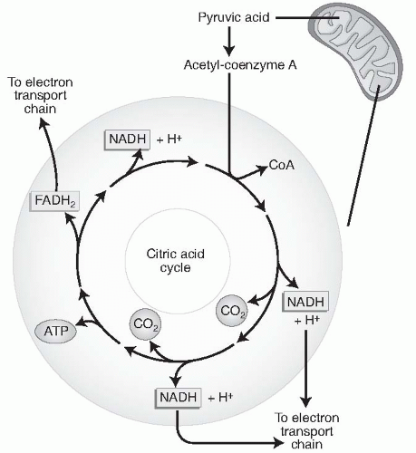
If oxygen is present (aerobic), the molecules of pyruvic acid move into the mitochondria where they enter the citric acid cycle, also called the Krebs cycle (
Fig. 1-4) and are converted by enzymes present there into a compound called acetyl coenzyme A (acetyl CoA). This process produces two more ATP molecules. Acetyl CoA is then enzymatically converted to carbon dioxide and hydrogen. The carbon dioxide diffuses out of the mitochondria and out of the cell, where it is picked up by the blood supplying that cell. It is then carried to the lungs and exhaled from the body. The hydrogen atoms remaining in the mitochondria begin the process of oxidative phosphorylation, during which they are combined with molecules of oxygen through an elaborate electron transport chain present in the mitochondrial membrane. The result of this process is to produce a tremendous amount of energy in the form of 36 ATP molecules. From the metabolism of one molecule of glucose, therefore, a total of 38 net ATP molecules are formed (36 from oxidative phosphorylation and 2 from glycolysis).
Oxidative Phosphorylation of Fatty Acids and Glycerol
The cell also uses free fatty acids and glycerol in oxidative phosphorylation to produce ATP. Glycerol is a three-carbon carbohydrate, which undergoes glycolysis in the cytoplasm and enters the Krebs cycle as acetyl CoA. Free fatty acids diffuse directly into the mitochondria where they are acted on by enzymes and transformed into acetyl CoA. This acetyl CoA also enters the Krebs cycle. The breakdown of one molecule of fat results in 463 molecules of ATP. Fat has a five times greater weight per mole than glucose. Thus, gram for gram, the metabolism of fat provides about three times as much ATP as glucose metabolism. Therefore, fat is a more efficient form of energy storage than carbohydrate.
Oxidative Phosphorylation of Amino Acids
Amino acids enter the mitochondria after removal of the nitrogen molecule (deamination). After deamination, amino acids enter the Krebs cycle at various points. Some, such as alanine, enter as pyruvic acid; others enter as later intermediates. Where the amino acids enter the Krebs cycle determines how many hydrogen atoms they add to the electron transfer chain and thus how many ATP molecules are synthesized.
Anaerobic Glycolysis
If oxygen is unavailable, the pyruvic acid produced by glycolysis does not enter the Krebs cycle, but combines with hydrogen in the cytoplasm to form lactic acid. Although the two molecules of ATP produced in the breakdown of one molecule of glucose to pyruvic acid are available to keep the cell alive, this is a wasteful use of glucose because it results in the loss of the other 36 molecules of ATP that would have been produced had pyruvic acid entered the Krebs cycle. This process can only continue for a short while before glucose is depleted.
The lactic acid produced by anaerobic glycolysis diffuses out of the cell and into the bloodstream. This can create a decrease in plasma pH (an increase in plasma acidity). With the return of oxygen, lactic acid will be reconverted to pyruvic acid, primarily in the liver, and the Krebs cycle will resume.
ENERGY USAGE
ATP formed in the mitochondria moves into the cytoplasm by a combination of simple and facilitated diffusion. When needed by the cell, ATP can be broken down rapidly into adenosine diphosphate (ADP) by enzymatic splitting of the bond between the last two phosphates. This results in the release of energy, which is used by the cell to perform its duties of solute transport, protein synthesis, reproduction, and movement.
Although it is through ATP synthesis and breakdown that energy is transferred in the cell, very little ATP is stored in the cell. Instead, energy is stored in the form of substrates for ATP—as carbohydrates, fats, proteins, and their metabolic products. The other essential component for ATP production, oxygen, is continually delivered to all cells by the combined efforts of the cardiovascular and respiratory systems.
CELL TYPES
Epithelial Cells
The tissue that lines most internal and external structures of the body is made up of epithelial cells. These cells are packed together, providing support for overlying structures. Epithelial tissue also acts as a protective barrier and a medium for absorption, secretion, and excretion. Examples of epithelial tissue include the skin (epidermis), the covering on all internal organs and tubules, the microvilli of the intestine, and the cilia lining the respiratory passageways. Glandular cells that secrete substances into ducts (exocrine glands) or into the bloodstream (endocrine glands) are made of epithelial tissue. Sensory organs also contain epithelial cells. Epithelial layers are usually one (simple epithelium) to two (stratified and pseudostratified epithelia) cells thick.
Connective Tissue Cells
Connective tissue is represented by many different cell types, including fibroblasts, adipose (fat) cells, mast cells, blood cells, and cells of the blood-forming organs. Connective tissue holds different tissues together by the accumulation of protein and gel-like substances secreted from the fibroblasts into the spaces surrounding the cells. Protein substances secreted include collagen, a thick, white fiber that acts to provide structural support; elastin, a stretchy protein that allows tissues to give when stretched; and reticular fibers, thin flexible strands that allow organs to accommodate increases in volume. Tissue gel consists primarily of hyaluronic acid, which intersperses throughout the interstitial spaces to retain water and provide support and protection.
Adipose tissue and endothelial cells provide nourishment and support for the fibroblasts. Mast cells contain granules filled with histamine and other vasoactive substances. Mast cell degranulation is an important step in initiating an inflammatory reaction.
Hematopoietic tissue is considered connective tissue. Hematopoietic tissue includes bone marrow, blood cells, and lymphatic tissue. The basement membrane found along the interface between connective tissue and an adjacent tissue is also considered a connective tissue layer. This membrane bonds, supports, and allows for tissue repair.
Muscle Cells
Muscle cells are highly differentiated (specialized) cells that have the ability to contract and cause movement or increased tension. Groups of muscle cells form one of the three types of muscle tissue: skeletal, smooth, or cardiac.
Muscle cells are composed of the proteins actin and myosin. Cross-bridges located between the actin and myosin connect and swing when stimulated in the proper sequence (see
Chapter 10). This causes the muscle as a whole to contract and do work or produce tension. All types of muscles require an increase in intracellular calcium to contract. Different muscles may use different sources of calcium and thus have slightly different methods of contraction stimulation.
Skeletal muscle is attached to bones by tendons. When stimulated by motor neuron impulses, skeletal muscle voluntarily contracts. Skeletal muscle uses calcium released from intracellular compartments to initiate contraction. Mature skeletal muscle does not undergo further cell division. Skeletal muscle may even be considered a paracrine endocrine organ because it secretes cytokines such as interleukin-8 (IL-8), a peptide that induces angiogenesis (new capillary formation).
Cardiac muscle, found in the heart, contracts spontaneously because of an intrinsic ability to depolarize and fire action potentials. Cardiac muscle is innervated by the nerves of the autonomic nervous system: the sympathetic and parasympathetic nerves. These inputs can increase or decrease the inherent rate or strength of cardiac contraction. Cardiac contraction involves entry of calcium into the muscle cell from the extracellular fluid and from an intracellular compartment, the sarcoplasmic reticulum. During embryogenesis, cardiac muscle cells become highly differentiated and do not undergo further cell division. Cardiac muscle is also an endocrine organ in that it secretes the hormone atrial natriuretic peptide (ANP) that acts on the kidney to participate in the control of blood volume.
Smooth muscle is found throughout the body, including the vascular system, the genitourinary tract, and all parts of the gut. Its function is often considered involuntary. Smooth muscle is innervated by the autonomic nervous system, which can increase or decrease the rate of contraction. When stretched, smooth muscle responds with an increase in contraction. Smooth muscle relies primarily on calcium entry from the extracellular fluid to initiate contraction. Mature smooth muscle cells can undergo cell division.


