The Pancreas and Diabetes Mellitus


The Pancreas and Diabetes Mellitus


The pancreas is a large, diffuse abdominal organ that functions as both an exocrine and an endocrine gland. Endocrine functions include the production and release of insulin, glucagon, and somatostatin. The secretion of digestive juices into the digestive tract is part of the exocrine function. In this chapter, both roles are presented, followed by a detailed description of diabetes mellitus, a condition in which the pancreatic hormone, insulin, is either ineffective or absent. Pancreatitis and pancreatic cancer are discussed briefly.


● Physiologic Concepts


EXOCRINE FUNCTIONS OF THE PANCREAS

The exocrine functions of the pancreas involve the synthesis and release of digestive enzymes and sodium bicarbonate from specialized cells of the pancreas called acini cells. The acini cells release their contents into the pancreatic duct. From the pancreatic duct, the enzymes and bicarbonate solution travel through the sphincter of Oddi into the first section of the small intestine, the duodenum. The pancreatic enzymes and bicarbonate solution both play important roles in the digestion and absorption of food in the small intestine.


Secretion of Pancreatic Enzymes

The secretion of the various pancreatic enzymes occurs primarily as a result of stimulation of the pancreas by cholecystokinin (CCK), a hormone released
from the small intestine. The stimulus for the release of CCK is the presence of a mixture of food particles entering the duodenum from the stomach. This food mixture coming from the stomach is called chyme. The pancreatic enzymes are secreted as inactive proenzymes that are activated when they reach the duodenum. The activated enzymes include:


  • trypsin, which is responsible for the digestion of proteins to amino acids


  • amylase, which is responsible for the digestion of carbohydrates to simple sugars


  • lipase, which is responsible for the digestion of fats to free fatty acids and monoglycerides


Secretion of Sodium Bicarbonate

Sodium bicarbonate is secreted from pancreatic ductal cells in response to a second small-intestine hormone, secretin. Secretin is released in response to the acidic chyme entering from the stomach. When delivered to the small intestine, sodium bicarbonate, which is a base, neutralizes acidic chyme. This function is essential because the digestive enzymes are inactivated in an acidic environment. Neutralization of the acid in the duodenum also protects this area against acid injury to the mucosal wall and subsequent development of ulcer.


ENDOCRINE FUNCTIONS OF THE PANCREAS

The endocrine functions of the pancreas involve the synthesis and release of the hormones insulin, glucagon, and somatostatin. These hormones are each produced by separate, specialized cells of the pancreas, called the islets of Langerhans.


Synthesis and Secretion of Insulin

The synthesis of insulin in the pancreas comes from the enzymatic cleavage of the molecule proinsulin, which itself is the cleavage product of an even larger preproinsulin molecule. Proinsulin is composed of an A peptide fragment connected to a B peptide fragment by a C peptide fragment and two disulfide bonds (Fig. 16-1). Enzymatic cleavage of the C peptide connections leaves the A and the B peptides connected to each other only through the two disulfide bonds. In this form, insulin circulates unbound in the plasma.

Insulin is released at a basal rate by the beta cells of the islets of Langerhans. A rise in blood glucose is the primary stimulus to increase insulin release above the baseline. Fasting blood glucose level is normally 80 to 90 mg/100 mL of blood. When blood glucose increases to more than 100 mg/100 mL of blood, insulin secretion from the pancreas increases rapidly and then returns to baseline in 2 to 3 hours. Insulin is the main hormone of the absorptive stage of digestion that occurs immediately after a meal. Insulin levels are low between meals.


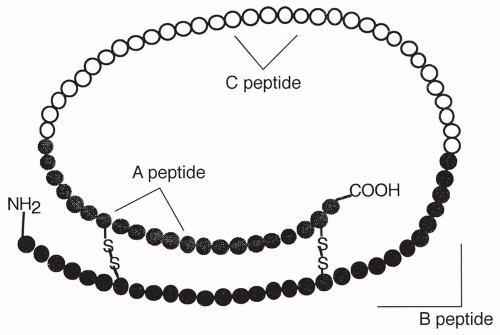
FIGURE 16-1 Proinsulin molecule.

Insulin circulates in the plasma and acts by binding to insulin receptors present in most cells of the body. Once bound, insulin works through a protein kinase messenger system to cause an increase in the number of glucose-transporter molecules present on the outside of the cell membrane. The glucose-transporter molecules, called glut-4 glucose transporters, are necessary for the facilitated diffusion of glucose into most cells. Once transported inside the cells, glucose can be used for immediate energy production through the Krebs cycle or it can be stored in the cell as glycogen, a glucose polymer, which is the storage form of glucose. When glucose is carried into the cell, it results in decreased blood levels of glucose, reducing further stimulation of insulin release. This cycle is an example of negative feedback, as shown in Figure 16-2.

Insulin release is also stimulated by amino acids and the digestive hormones (i.e., CCK, secretin, and glucose-dependent insulinotropic polypeptide [GIP]; see Chapter 15). The autonomic nervous system also stimulates insulin release by means of parasympathetic nerves to the pancreas. Both the release of GIP and the activation of the autonomic nervous system occur when one starts eating, resulting in a release of insulin at the beginning of a meal, even before glucose is absorbed. Sympathetic stimulation of the pancreas decreases insulin release.

Insulin is the major anabolic (building) hormone of the body and has a variety of other effects besides stimulating glucose transport. It also increases amino acid transport into cells, stimulates protein synthesis, and inhibits the breakdown of fat, protein, and glycogen stores. Insulin also inhibits gluconeogenesis, the synthesis of a new form of glucose, by the liver. In summary,
insulin serves to provide glucose to our cells, build protein, and maintain low plasma glucose levels.


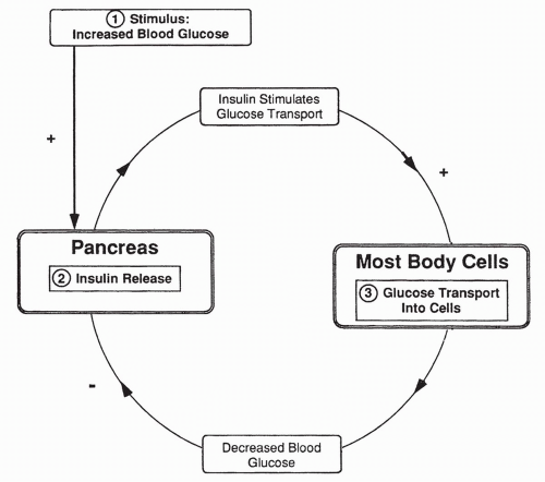
FIGURE 16-2 Feedback cycle demonstrating the effect of decreased blood glucose on insulin release.


The Brain, Glucose, and Insulin

Unlike most other cells, brain cells do not require insulin for glucose entry. Also unlike other cells that may use free fatty acids or amino acids for energy, brain cells must use only glucose or glycogen to meet their energy demands and drive their cellular functions. In other words, brain cells are obligate users of glucose and glycogen. This means that gluconeogenesis by the liver is important; if glucose were not produced between meals by the liver, the brain would have no usable energy source during that time.


Secretion of Glucagon

Glucagon is a protein hormone released from the alpha cells of the islets of Langerhans in response to low blood glucose levels and increased plasma amino acids. Glucagon is primarily a hormone of the postabsorptive stage of digestion
that occurs during fasting periods in between meals. Its functions are mainly catabolic (breaking down). In most respects, the functions of glucagon are opposite to that of insulin. For example, glucagon acts as an insulin antagonist by inhibiting glucose movement into cells. Glucagon also stimulates liver gluconeogenesis and causes the breakdown of stored glycogen to be used as an energy source instead of glucose. Glucagon stimulates the breakdown of fats and the release of free fatty acids into the bloodstream so they may be used as an energy source instead of glucose. These functions serve to increase blood glucose levels. The release of glucagon by the pancreas is stimulated by sympathetic nerves.


Secretion of Somatostatin

Somatostatin is secreted by delta cells of the islets of Langerhans. Somatostatin is also called growth hormone-inhibiting hormone and is released by the hypothalamus as well. Somatostatin from the hypothalamus inhibits the release of growth hormone from the anterior pituitary. Somatostatin from the pancreas appears to have a minimal effect on the release of growth hormone from the pituitary. It rather controls metabolism by inhibiting the secretion of insulin and glucagon. The exact function of somatostatin is otherwise unclear.


Counter-Regulatory Hormones

Glucose levels may also be affected by catecholamines, growth hormone, or glucocorticoid. These are known as counter-regulatory hormones and they facilitate glucose regulation when glucose intake is decreased or glucose stores are depleted. Table 16-1 includes a brief description of each.


TESTS OF PANCREATIC FUNCTION


Fasting Plasma Glucose

Plasma glucose levels provide instantaneous data about the glucose level at that moment. Measurement of plasma glucose above 126 mg/100 mL (corresponding to fasting blood glucose of 110 mg/100 mL) on more than one occasion is diagnostic of diabetes mellitus. Plasma glucose levels greater than 110 mg/100 mL indicate insulin resistance. Nonfasting plasma glucose level greater than 200 mg/100 mL with symptoms of polyurea, polydipsia, and polyphagia is also diagnostic of diabetes.


Urine Glucose Tests

Glucose in the urine may or may not be indicative of diabetes. Likewise, the absence of glucose in the urine cannot be used to discount diabetes. Under most conditions, however, glucose is not present in the urine of healthy, nonpregnant individuals and further assessment is necessary when it is present.


TABLE 16-1 Counter-Regulatory Hormones


Hormone


Description


Epinephrine


  • Released from adrenal medulla during stressful periods


  • Causes glycogenolysis in the liver


  • Inhibits insulin release from beta cells


  • Increases breakdown of muscle glycogen stores which in turn frees up glucose to be used by other organs


  • Lipolytic effect on adipose cells which mobilizes fatty acids for energy use


  • Facilitates homeostasis during a hypoglycemic episode


Growth hormone


  • Increases protein synthesis


  • Mobilizes fatty acids from adipose tissue


  • Decreases cellular uptake and use of insulin


  • Antagonizes effects of insulin


  • Decreases peripheral use of glucose


Glucocorticoid hormones


  • Essential for survival during starvation


  • Stimulate gluconeogenesis by the liver


  • Moderately decrease tissue use of glucose


Glycosylated Hemoglobin

Throughout the 120-day life span of the red blood cell, hemoglobin slowly and irreversibly becomes glycosylated (glucose bound). Normally, approximately 4% to 6% of red blood cell hemoglobin is glycosylated. If there is chronic hyperglycemia, the level of glycosylated hemoglobin increases. Patients with poorly controlled diabetes show the highest level of glycosylated hemoglobin, which may be greater than 10%. The particular hemoglobin most often measured and reported is glycohemoglobin A1c (HbA1c). Measurement of HbA1c is important because it offers an indication of how well controlled the blood glucose has been over the previous 2 to 4 months.


1,5-Anhydroglucitol (1,5-AG)

A relatively new measurement offers hope for improving therapeutic management of glucose control. 1,5-AG offers information about glucose control over days to weeks as opposed to the 2 to 4 month period of HbA1C.


Serum Amylase and Lipase

Amylase is a pancreatic enzyme. Its increased concentration in the serum suggests pancreatic acinar damage and pancreatic destruction. Lipase is a digestive
enzyme secreted solely by the pancreas and elevated serum levels indicate damage to pancreatic acinar cells.


● Pathophysiologic Concepts


HYPOGLYCEMIA

Hypoglycemia occurs when the blood glucose level falls below 50 mg/100 mL of blood. Hypoglycemia is caused by an imbalance between glucose production and utilization. It can be caused by fasting or, especially, fasting coupled with exercise, because exercise increases the usage of glucose by skeletal muscle. Most commonly, hypoglycemia is caused by an insulin overdose in an insulin-dependent diabetic.

The brain relies on glucose as its main energy source and because it cannot synthesize or store glucose, it is dependent on circulating blood glucose to maintain normal functioning. Hypoglycemia results in many symptoms of altered central nervous system (CNS) functioning, including confusion, irritability, seizure, and coma. Hypoglycemia can cause headache, as a result of alteration of cerebral blood flow, and changes in water balance. Systemically, hypoglycemia causes activation of the sympathetic nervous system, stimulating hunger, nervousness, sweating, and tachycardia. Anxiety levels increase due to the diabetic being shaky and agitated.


HYPERGLYCEMIA

Hyperglycemia is defined as a condition when plasma glucose level is higher than the normal (fasting range of 126 mg/100 mL of blood). Hyperglycemia is usually caused by insulin deficiency, as seen in type 1 diabetes, or as a result of decreased cellular responsiveness to insulin, as seen in type 2 diabetes (the types of diabetes are discussed in the following section). Hypercortisolemia, which occurs in Cushing syndrome and in response to chronic stress, can cause hyperglycemia by the stimulation of liver gluconeogenesis. Acute conditions of elevated thyroid hormone, prolactin, and growth hormone all increase blood glucose as well. Prolonged high levels of these hormones, especially growth hormone, are considered diabetogenic (producing diabetes) because they overstimulate insulin release by beta cells of the pancreas, leading to an eventual decrease in the cellular response to insulin. Sympathetic nervous stimulation and epinephrine released from the adrenal gland also raise plasma glucose levels, especially during periods of stress. The catecholamines epinephrine and norepinephrine inhibit insulin secretion, increase the breakdown of stored fats, and promote the use of glycogen for energy. By these mechanisms, the catecholamines make a variety of alternative energy sources available for the body to use instead of glucose, thereby raising plasma glucose levels and increasing its availability for use by the brain.


● Conditions of Disease or Injury


DIABETES MELLITUS

Diabetes is a Greek word that means “to siphon or pass through.” Mellitus is a Latin word meaning honey or sweet. The disease diabetes mellitus is one in which an individual siphons large volumes of urine with a high glucose level. It is a disease of hyperglycemia characterized by the absolute lack of insulin or a relative lack of cellular insensitivity to insulin. Based on recent epidemiological evidence, the number of people afflicted with diabetes around the globe, currently nearly 200 million, is expected to increase to over 330 million by the year 2025. Reasons for the increase include longer life expectancy and higher population growth coupled with increased rates of obesity associated with urbanization and reliance on processed foods. In the United States, of the 18.2 million persons with diabetes (6.3% of the population), nearly one third are unaware that they have the disease. It is further projected that 57 million people in the United States have prediabetes.

Tests used to diagnose diabetes include the fasting plasma glucose (FPG) test and the oral glucose tolerance test (OGTT). The American Diabetes Association recommends the FPG test because it is faster, easier to perform, and less expensive than the OGTT. An FPG level between 100 and 125 mg/dL is indicative of prediabetes, and an FPG level of 126 mg/dL or more is considered as frank diabetes. For the OGTT, a person’s blood glucose is measured after a fast and 2 hours after drinking a glucose-rich beverage. A 2-hour OGTT between 140 and 199 mg/dL indicates prediabetes; a level of 200 mg/dL or higher indicates diabetes. Providing a range of values indicative of prediabetes allows for earlier intervention in patients at risk of developing frank diabetes. Early intervention is extremely important because, at the time of diagnosis of type 2 diabetes, 20% of patients already have retinal damage, 8% have renal dysfunction, and 9% have neurologic symptoms. Even if the FPG is within normal limits, an OGTT should be conducted if the individual has risk factors for prediabetes. Box 16-1 includes risk factors identified by the American Diabetes Association.


Types of Diabetes Mellitus

In 2007, the American Diabetes Association updated the classification of diabetes and abnormal glucose tolerance to include five types of diabetes. The new criteria are based on the etiology of the disease rather than the presentation. The five types include type 1, type 2, gestational diabetes, other types of diabetes, and prediabetes (Table 16-2). Types 1, 2, and gestational diabetes are discussed in the following sections. Other specific types of diabetes include those due to pancreatic trauma, neoplasm, or diseases characterized by other endocrine disorders, for example, Cushing disease (see Chapter 9).


TABLE 16-2 Diabetes Mellitus: A Classification Scheme


Type


Characteristics


Etiology


Treatment


Type 1


Absolute lack of insulin


Autoimmune


Insulin


Type 2


Insulin insensitivity and insulinsecreting deficiency


Obesity, genetics


Diet, exercise, hypoglycemic agents, transporter-stimulating drugs


Gestational


First diagnosed during pregnancy


Increased metabolic demands, family history of diabetes, decreased insulin sensitivity


Diet, hypoglycemic agents, insulin


Other types


Other specific cause


Dependent on specific cause


Dependent on cause


Prediabetes


Above normal FPG or OGTT, but not abnormal enough to meet criteria for diabetes


Insulin resistance


Regular monitoring


Latent autoimmune diabetes of adulthood (LADA) is a type of diabetes that is frequently misdiagnosed because it has characteristics of both type 1 and type 2. The body mistakenly responds to insulin-producing beta cells of the pancreas as it does to foreign cells. This mistaken identity causes the body to attack and destroy the beta cells. They typically present with an insidious onset of hyperglycemia and no evidence of insulin resistance. Because beta cell destruction is fairly slow, the need for insulin occurs much later than with type 1 diabetes, but sooner than with type 2 diabetes.


Type 1 Diabetes Mellitus

Hyperglycemia caused by an absolute lack of insulin is known as type 1 diabetes mellitus. Previously, this type of diabetes has been referred to as insulin-dependent diabetes mellitus (IDDM) because individuals who have this disease must receive insulin replacement. Type 1 diabetes is most commonly seen in nonobese individuals less than 30 years old and occurs in a slightly higher proportion of males than females. Because the incidence of type 1 diabetes peaks in the early teens, in the past it was referred to as juvenile diabetes. However, type 1 diabetes mellitus can occur at any age. See page C13 for illustrations and further explanation.


Causes of Type 1 Diabetes.

Type 1 diabetes results from autoimmune destruction of the beta cells of the islets of Langerhans. It appears that individuals who have a genetic tendency to develop this disease experience an environmental trigger that initiates the autoimmune process. Examples of possible triggers include viral infections such as mumps, rubella, or chronic cytomegalovirus (CMV). It also has been suggested that exposure to certain drugs or toxins may trigger an attack. Because type 1 diabetes develops over several years, there is often no clear stimulating event. Antibodies to islets of Langerhans cells are present in most individuals at the time of diagnosis of type 1 diabetes.

Why an individual develops antibodies against the islet of Langerhans cells in response to a triggering event is unknown. One mechanism may be that the environmental agent antigenically changes the cells such that they stimulate the production of autoantibodies. It is also possible that individuals who develop type 1 diabetes mellitus share antigenic similarities between their pancreatic beta cells and certain triggering microorganisms or drugs. In the course of responding to a virus or drug, the immune system may fail to distinguish the pancreatic cells as self.


Genetic Tendency for Type 1 Diabetes Mellitus.

There appears to be a genetic tendency for individuals to develop type 1 diabetes mellitus. Certain individuals appear to have diabetogenic genes, meaning a genetic profile that predisposes them to type 1 diabetes (or possibly any autoimmune disease). Genetic loci that pass an inherited tendency for type
1 diabetes are part of the histocompatibility complex genes (see Chapter 4). The histocompatibility complex controls the recognition of self-antigens by the immune system; loss of self-tolerance is core to developing autoantibodies. The histocompatibility genes are primarily coded on chromosome 6. Another specific insulin-related gene on chromosome 11 has been implicated in the development of type 1 diabetes through its effects on beta cell development and replication. Siblings of individuals who have type 1 diabetes and children of a parent who has type 1 diabetes have an increased risk of developing the disease compared with those without an affected first-degree relative. In clinical studies, nonsymptomatic siblings show a higher incidence (2% to 4%) of antibodies against pancreatic beta cells compared to those who do not have a first-degree relative with diabetes; the earlier the onset of antibodies and the higher the level, the greater the likelihood of those siblings developing the disease later in life.


Characteristics of Type 1 Diabetes.

Individuals who have type 1 diabetes show normal glucose handling before disease onset. In the past, it was thought that type 1 disease developed suddenly and with little warning. Currently, however, it is thought that type 1 diabetes usually develops slowly over the course of many years, with the presence of autoantibodies against the beta cells and their steady destruction occurring well in advance of diagnosis.

Only gold members can continue reading. Log In or Register to continue

Stay updated, free articles. Join our Telegram channel

Jun 17, 2016 | Posted by in PATHOLOGY & LABORATORY MEDICINE | Comments Off on The Pancreas and Diabetes Mellitus

Full access? Get Clinical Tree

Get Clinical Tree app for offline access