The Endocrine System


The Endocrine System


The endocrine system, along with the nervous system, is a chemical communication and coordination system. There are three components to the endocrine system: endocrine glands that secrete chemical messengers into the bloodstream; the chemical messengers themselves, called hormones; and the target cells or organs that respond to the hormones (see Table 9-1). The major functions of the endocrine system include:


  • Regulation of digestion


  • Storage and use of nutrients


  • Electrolyte and water metabolism


  • Growth and development


  • Reproduction


  • Synthesis, storage, and secretion of hormones.


● Physiologic Concepts


ENDOCRINE GLANDS

Endocrine glands are organs that synthesize, store, and secrete hormones into the bloodstream. There are many endocrine glands in the body, including the hypothalamus, pituitary gland, adrenal glands, gonads, pineal gland, thymus, pancreas, thyroid, and parathyroid. Other hormone-secreting organs include the kidneys (erythropoietin), heart (atrial natriuretic factor), and gut (peptide hormones). The endocrine glands reviewed in this chapter are the hypothalamus, the anterior and posterior pituitary glands, and the glands that function as target organs for the pituitary hormones.


TABLE 9-1 Endocrine Glands, Hormones, and Target Cells


Gland


Hormone(s)


Target Cells


Hypothalamus


Thyrotropin-releasing hormone (TRH)


Anterior pituitary


Corticotropin-releasing hormone (CRH)


Anterior pituitary


Gonadotropin-releasing hormone (GnRH)


Anterior pituitary


Growth hormone-releasing hormone (GHRH)


Anterior pituitary


Somatostatin


Pituitary, pancreas, GI tract


Dopamine


Anterior pituitary


Anterior pituitary (adenohypophysis)


Growth hormone (GH)


Systemic


Adrenocorticotropic hormone (ACTH)


Adrenal cortex


Thyroid-stimulating hormone (TSH)


Thyroid


Follicle-stimulating hormone (FSH)


Ovaries, testes


Luteinizing hormone (LH)


Ovaries


Prolactin


Mammary glands


Melanocyte-stimulating hormone


Skin, hair


Posterior pituitary (neurohypophysis)


Antidiuretic hormone (ADH)


Kidney


Oxytocin


Breast, uterus, testes


Thyroid gland


Thyroxine (T4)


Systemic


Triiodothyronine (T3)


Systemic


Thyrocalcitonin (calcitonin)


Bone


Parathyroid gland


Parathyroid hormone (PTH)


Bone, kidney, intestine


Adrenal glands (cortex)


Mineralocorticosteriods (mainly aldosterone)


Kidneys


Glucocorticoids (mainly cortisol)


Most tissues have receptors


Adrenal androgens


Gonads


Adrenal glands (medulla)


Epinephrine


Cardiac muscle, smooth muscle, glands


Norepinephrine


Sympathetic nervous system


Pancreas


Insulin


Muscles, tissues


Glucagon


Liver


Somatostatin


Gonads (males)


Androgens (testosterone)


Gonads, muscle tissue


Gonads (females)


Inhibin


Ovaries, pituitary, placenta


Oestrogens (estrogen)


Breasts, uterus


Corpus luteum


Testosterone


Endometrium


Progesterone


Endometrium


Placenta


Human chorionic gonadotropin (HCG)


Endometrium


Human placental lactogen (hPL)


Placenta


Progesterone


Placenta


Corticotropin-releasing hormone (CRH)


Placenta


Kidneys


Calcitriol (1,25-dihydroxy-vitamin D)


Intestine


Renin


Kidneys


Erythropoietin (EPO)


Bone marrow


Gastrointestinal tract


Gastrin


Stomach


Enterogastrone


Stomach


Secretin


Liver, pancreas


Pancreozymin


Pancreas


Ghrelin


Pituitary


Cholecystokinin (CCK)


Gallbladder


Liver


Insulin-like growth factor


Nerve, muscle, other cells


Angiotensinogen


Vascular, brain, kidneys, adrenals


Thrombopoietin


Bone marrow, platelets


Brain


Peptide hormones


Brain, nervous system


Heart


Atrial-natriuretic peptide (ANP)


Heart


Adipose tissue


Leptin


Brain, nervous cells


Resistin


Adipose cells


Bones


Osteocalcin


Pancreas, fat cells


Skin


Calciferol (Vitamin D)


Kidneys, intestines, bones


The Hypothalamus

The hypothalamus is a small area of the brain located in the section of the forebrain called the diencephalon. The hypothalamus is a neural and an endocrine organ functioning to maintain homeostasis in the body’s internal environment. The hypothalamus is also essential in controlling behavior and allowing appropriate responses to multiple incoming stimuli. It continually receives information from the central and peripheral nervous systems concerning temperature, pain, pleasure, feeding, hunger, body mass, and metabolic status. It also receives input from other hormones of the body and receives neural extensions from other areas of the brain.

The hypothalamus, in turn, responds to all the incoming stimuli by sending neural projections throughout the brain and by synthesizing and secreting its own hormones. Nerve cell bodies in the ventral hypothalamus synthesize several hormones and send them in axon projections to be released into the blood and delivered to the anterior pituitary gland. Other nerve cell bodies in the hypothalamus synthesize hormones that are sent down via axon projections to the posterior pituitary, where they are stored until they are eventually released into the bloodstream. Regulatory hormones include thyrotropin-releasing hormone (TRH), gonadotropin-releasing hormone (GnRH), growth hormone-releasing hormone (GHRH), growth hormone-inhibiting hormone (GHIH) somatostatin, corticotropin-releasing hormone (CRH), prolactin-inhibiting hormone (PIH), and melanocyte-inhibiting hormone (MIH). The two routes by which the hypothalamus controls hormone release by the anterior and posterior pituitary are shown in Figure 9-1.


The Anterior Pituitary

The anterior pituitary, also called the adenohypophysis, is composed of nonneural tissue. It is anatomically separate from the hypothalamus, but functionally connected to it through its blood supply. The anterior pituitary receives its blood through venous drainage from the hypothalamus. When blood flowing in a vein breaks into another capillary network instead of flowing back to the vena cava,
the system is called a portal venous system. Thus, the hypothalamus and the anterior pituitary are connected by the hypothalamic-anterior pituitary portal blood flow system. Because this blood has already been used by the hypothalamus, it is poorly oxygenated but rich in hormonal messages put out by the hypothalamus into the median eminence (see later). The anterior pituitary is, therefore, a major target organ for hypothalamic hormones and responds to these hormones with the release of its own hormones that stimulate various glands or produces a primary effect on cells with mediation of other endocrine glands.


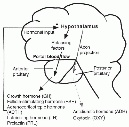
FIGURE 9-1 The hypothalamic-pituitary system. The hypothalamus is connected through the blood to the anterior pituitary while the posterior pituitary is a neural outgrowth.


The Posterior Pituitary

The posterior pituitary, also called the neurohypophysis, is true neural tissue derived embryologically from the hypothalamus. There are three parts to the posterior pituitary: the median eminence (sometimes considered hypothalamic tissue), into which the hypothalamus secretes the anterior pituitary-releasing hormones; the infundibular stem connecting the hypothalamus with the posterior pituitary; and the infundibular process, which is the terminal end of the posterior pituitary.

Nerve cell bodies in the supraoptic and paraventricular nuclei of the hypothalamus synthesize two hormones: antidiuretic hormone (ADH), also
called vasopressin, and oxytocin. The hypothalamus sends these hormones in axon projections through the infundibular stem to the infundibular process. They are stored there until the hypothalamus stimulates them to be released into the general circulation. Thus, the hormones released by the posterior pituitary are hypothalamic in origin and depend on the hypothalamus for their release.


Target Glands

The third group of endocrine glands discussed in this chapter consists of those outside the brain that respond to the anterior and posterior pituitary hormones with the release of their own hormones. These glands are the target organs of the pituitary hormones and include the thyroid gland, the adrenal gland, and the testes and ovaries. The pancreas, which secretes insulin, is also an endocrine gland and is discussed in Chapter 16.


HORMONES

A hormone is a chemical messenger released by an endocrine gland into the circulation. Once released, a hormone travels in the bloodstream and affects only cells in the body that have receptors (binding sites) specific to it. Cells that respond to a particular hormone are called target cells for that hormone. Many hormones may be necessary to initiate a single function and conversely, a single hormone may influence many different tissues in the body.

Typically, a hormone is released in bursts from an endocrine gland in a pattern that often follows an inherent daily (diurnal) rhythm. The burst of hormone release can be increased or decreased above or below baseline level by various inputs to the gland. Inputs that affect hormone release involve: (1) stimulation by another hormone or neurotransmitter, or (2) stimulation caused by a decrease or increase in a certain ion or nutrient. Examples of hormones that cause an increase or decrease in another hormone’s release include all the hypothalamic hormones affecting the anterior pituitary. Examples of neurotransmitters affecting a hormone’s release include the release of insulin in response to epinephrine and norepinephrine stimulation. Ions that influence the release of a hormone include calcium ion’s effect on parathyroid hormone, and sodium ion’s effect on aldosterone. Nutrients that affect the release of hormones include the amino acids that stimulate the release of insulin and growth hormone (GH). Frequently, one endocrine gland is stimulated simultaneously by several different inputs.

There are three broad categories of hormones: peptide, steroid, and amino acid. Most hormones, including all the hypothalamic and pituitary hormones, are peptide hormones. The steroid hormones are made from cholesterol and are soluble across the cell membrane. The amino acid hormones are made from
the amino acid tyrosine. A fourth category, the fatty acid derivatives, includes the retinoids and eicosanoids.


Peptide Hormones

Peptide hormones range in size from a few amino acids to relatively large protein complexes. Peptide hormones circulate in the plasma to their target organs and exert their effects by binding to specific receptors present on the outside of target cell membranes. By binding to its receptor, a protein hormone changes the cell’s permeability to water, electrolytes, or organic molecules such as glucose, or causes the activation of intracellular messengers, which then causes enzyme activation or protein synthesis. Examples of intracellular messengers include the G proteins, which many protein hormones first activate during receptor binding, and the second messengers such as cyclic adenosine monophosphate (cyclic AMP) and calcium, which are subsequently activated by the G proteins. Peptide hormones include hypothalamic-releasing and -inhibiting hormones and factors, anterior pituitary protein hormones, posterior pituitary hormones, hormones of digestion and metabolism (Chapters 15, 16), hormones of blood pressure and electrolyte balance (Chapters 13, 19), hormone for red blood cell development (Chapters 12, 19), and hormone to modulate stress and pain (Chapters 6, 8, 9).


Steroid Hormones

Steroid hormones are cholesterol-based, lipid-soluble molecules produced by the adrenal cortex and the sex organs. Because steroid hormones are lipid-soluble, they can cross the cell membrane and bind to receptors or carriers inside the cell. Once inside a cell, the steroid hormone travels to the cell nucleus, where it influences the cell by affecting DNA replication, transcription of DNA into RNA, or translation of RNA into proteins. Because this is a lengthy process, steroid hormone responses typically take longer to occur than responses caused by nonsteroid hormones. Steroid hormones are discussed in this chapter and in Chapter 20. Steroid hormones include gonadal hormones, estrogens, progesterones, androgens, and hormones of the adrenal cortex.


Amine Hormones

The amine hormones are derivatives of the amino acid tyrosine and include thyroid hormone (TH) and the catecholamines (epinephrine, norepinephrine, and dopamine). Epinephrine, norepinephrine, and dopamine also act as neurotransmitters in the central and peripheral nervous systems. The catecholamine hormones travel in the blood to their target cell and bind to the plasma membrane at specific receptor sites. Binding of catecholamine activates the cyclic AMP second messenger system and alters enzyme activity or
membrane permeability. TH travels in the blood mostly bound to carrier proteins with a smaller amount circulating free. Once at the target cell, free TH crosses the cell membrane and binds to the nuclear DNA, directly affecting DNA transcription. Therefore, free hormone, although lesser in quantity, is the active hormone. Amine hormones include aldosterone, glucocorticoids, androgens, and estrogens.


FEEDBACK

In the endocrine system, feedback refers to the response of a target tissue after stimulation by a specific hormone which then influences the continued release of that hormone. Each hormone is stimulated to be released by a specific signal. Once released, a hormone affects its target organ, causing a response that usually reduces further hormone release. This type of feedback, shown in Figure 9-2, is called negative feedback, and allows tight control over hormone levels. Positive feedback is uncommon and occurs when the response by a target
tissue to hormonal stimulation increases the further release of that hormone. The feedback mechanism is a series of reactions that work to achieve homeostasis by neuroregulatory mechanisms. The central nervous system (CNS) receives input that is transmitted to the hypothalamus. The hypothalamus then produces and releases either releasing or inhibiting factors that are transported to the pituitary. In the pituitary, releasing or inhibiting factors release or inhibit specific hormones. The anterior pituitary responds by controlling secretion of hormones from target organs or tissues.


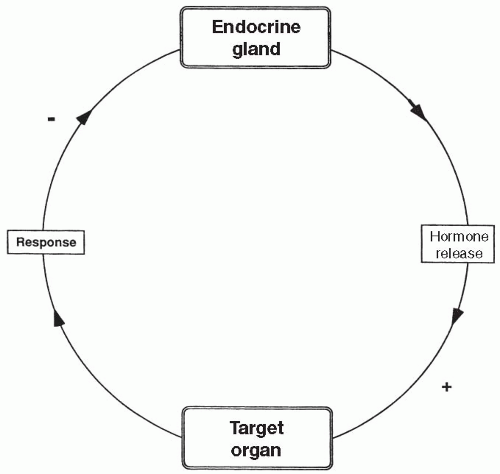
FIGURE 9-2 A typical negative feedback cycle in which an endocrine gland releases a hormone, which then stimulates (+) its target organ to respond in such a way that further secretion of the hormone by the endocrine gland is reduced (−).


FACTORS CONTROLLING HORMONE SECRETION


Factors Controlling Anterior Pituitary Hormone Secretion

The stimuli that control the secretion of the pituitary hormones (except melanocyte-stimulating hormone) are the hormones secreted by the hypothalamus that travel in the portal blood to the anterior pituitary. These hormones are hypothalamic-releasing or hypothalamic-inhibiting hormones, depending on whether they increase or decrease the release of the pituitary hormone they control. When a hypothalamic-releasing hormone is secreted, its corresponding anterior pituitary hormone is released. When a hypothalamic-inhibiting hormone is secreted, it inhibits synthesis and release of the anterior pituitary hormone over which it has control. Once secreted, the pituitary hormones act to stimulate another target organ or cell to perform a function or release a hormone of its own.

The pituitary hormone and the subsequent response to it by its target organ may feed back on the hypothalamus to decrease further release of the hypothalamic hormone. The target organ response may also inhibit further release of the pituitary hormone.


Factors Controlling Hypothalamic Hormone Secretion

For the hypothalamic-pituitary hormonal system, the hypothalamus ultimately determines whether a hormone will be secreted. The hypothalamic-releasing or -inhibiting hormones are secreted at a baseline level that can be increased or decreased as a result of the integration of many neural inputs to the hypothalamus. The inputs are related to stress, pain, body weight, temperature, emotions, and various hormones released by target organs. All these influences can be excitatory or inhibitory for each releasing or inhibiting hormone.


TARGET ORGAN HORMONES


Thyroid Hormone

TH is an amine hormone synthesized and released from the thyroid gland. It is made when one or two iodine molecules are joined to a large glycoprotein
called thyroglobulin, which is synthesized in the thyroid gland and contains the amino acid tyrosine. These iodine-containing complexes are called iodotyrosines. Two iodotyrosines then combine to form two types of circulating TH, called T3 (triiodothyronine) and T4 (thyroxine). T3 and T4 differ in the total number of iodine molecules they contain (three for T3 and four for T4). Approximately 90% of the TH released into the bloodstream is T4, but T3 is physiologically more potent. A diet adequate in iodine and protein is necessary for T3 and T4 to be produced in adequate amounts. TH is stored in the thyroid gland as a colloid compound until needed. In passage through the liver and kidney, most T4 is converted to T3. T3 and T4 are carried to their target cells in the blood bound to a plasma protein, but enter the cell as free hormone. T3 and T4 collectively are referred to as TH.


Effects of Thyroid Hormone

Target cells for TH include almost all cells of the body. Effects of TH are listed below.


  • Stimulates metabolic rate of all target cells by increasing the metabolism of protein, fat, and carbohydrate (primary function).


  • Stimulates the rate of the sodium-potassium pump in its target cells.


  • Increases utilization of energy by the cells, thereby increasing basal metabolic rate (BMR), burning calories, and increasing heat production by each cell (as a result of the two effects listed above).


  • Increases sensitivity of target cells to catecholamines, thus increasing heart rate and causing heightened emotional responsiveness.


  • Increases rate of depolarization of skeletal muscle, which increases the speed of skeletal muscle contractions, often leading to a fine tremor.


  • Essential for normal growth and development of all cells of the body and is required for the function of GH.


  • Increases red blood cell production.


Factors Controlling Thyroid Hormone Secretion

The stimulus for the secretion of TH is thyroid-stimulating hormone (TSH), released into the bloodstream from the anterior pituitary. The stimulus for the release of TSH is thyroid-releasing hormone (TRH), secreted from the hypothalamus into the portal bloodstream. TH appears to act in a negative feedback manner on the hypothalamus, to decrease the further release of TRH, and on the pituitary, to decrease the release of TSH. TSH may also act on the hypothalamus to decrease further release of TRH.


Factors Controlling Thyroid-Releasing Hormone Secretion

The stimuli responsible for increasing TRH secretion include exposure of the body to cold temperature, physical and perhaps psychological stress, and low
levels of TH. When the secretion of TRH is stimulated by cold temperature, the result is an increase in TH, which increases BMR, thereby increasing body heat and reducing the demand for a further increase in TRH (Fig. 9-3). This is an example of negative feedback.


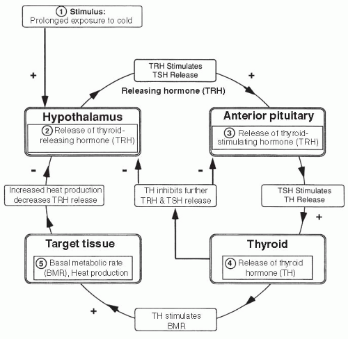
FIGURE 9-3 Feedback: thyroid hormone.


Glucocorticoids

Glucocorticoids are steroid hormones released from the cortex (outer layer) of the adrenal gland that affect many aspects of metabolism, especially glucose metabolism. In humans, the main glucocorticoid is cortisol. The glucocorticoids also affect many other systems of the body, including the cardiovascular and immune systems. Glucocorticoids are released in a diurnal (daily) manner, peaking in the early morning hours.


Effects of the Glucocorticoids

The major effects of glucocorticoids are listed below. Many of these glucocorticoid effects are essential in times of trauma and stress. They allow one to survive blood loss, periods of hunger or starvation, or prolonged exposure to environmental extremes.


  • Increase the level of blood glucose by stimulating gluconeogenesis (conversion in the liver of fats and proteins into glucose).


  • Increase blood glucose levels by stimulating muscle, adipose (fat), and lymphatic tissues to use free fatty acids for energy instead of glucose.


  • Stimulate protein breakdown and inhibit protein synthesis in all body cells.


  • Stimulate hunger, promote fat buildup in the trunk and face, and inhibit growth by suppressing GH and antagonizing the effects of GH on protein synthesis.


  • Increase the effect of GH on adipose tissue and increase the effect of TH on its target tissues.


  • Increase the effects of the catecholamines, causing increased heart rate and blood pressure.


  • Nonmetabolic effect that occurs with high circulating levels of cortisol include inhibition of immune and inflammatory functions by blocking almost every component of the immune and inflammatory responses including depressed cytotoxic T-cell function and suppression of the production, release, and activation of many chemical mediators of inflammation, including interleukins, prostaglandins, and histamine. Levels of cortisol high enough to inhibit immune and inflammatory function may be reached with pharmacologic administration of cortisol for immunosuppression, with tumors of the adrenal gland, or with long-term stress.


  • Major effect on emotional stability and mood.


Factors Controlling Glucocorticoid Release

Glucocorticoids are released from the adrenal gland in response to circulating adrenocorticotropic hormone (ACTH) from the anterior pituitary. ACTH is released in response to CRH carried in the portal blood from the hypothalamus. CRH also stimulates the release of endorphins by the anterior pituitary and perhaps elsewhere. When released, glucocorticoids feed back on the hypothalamus and on the anterior pituitary to decrease the further release of CRH and ACTH, respectively.


Factors Controlling Corticotropin-Releasing Hormone

CRH is secreted from the hypothalamus in a diurnal pattern that sets the subsequent release pattern of ACTH and cortisol. Stimuli for an increase in CRH include stress, hypoglycemia (low blood glucose), and decreased circulating levels of glucocorticoids. The feedback cycle of CRH release in response to hypoglycemia is shown in Figure 9-4.


Other Effects of Adrenocorticotropic Hormone

Adrenal androgens are released in response to ACTH stimulation of the adrenal gland. Adrenal androgens are the primary source of androgens in women and
children. High levels of ACTH can result in masculinization of women and children. ACTH is similar in structure to another anterior pituitary hormone, melanin-stimulating hormone (MSH), which causes the cells of the skin to produce the tanning substance melanin. High levels of ACTH can have crossover effects on the skin and cause bronzing. A limited amount of ACTH appears essential for the synthesis of another adrenal cortical hormone, aldosterone. Without aldosterone, salt wasting and death occur.


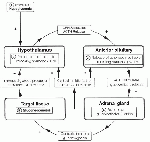
FIGURE 9-4 Feedback: glucocorticoids.


Growth Hormone

GH, also called somatotropin, is a protein hormone released in a diurnal pattern over 24 hours. Approximately 70% of daily secretion occurs in a burst 1 to 4 hours after the onset of sleep. Accelerated GH release occurs during puberty and pregnancy.


Effects of Growth Hormone

GH effects are listed below.


  • Increases protein synthesis in all cells of the body, especially muscle cells.


  • Stimulates the growth of cartilage and activity of osteoblasts, the boneproducing cells of the body.


  • Essential for longitudinal bone growth and for the continual remodeling of bone which occurs throughout life. Effects of GH on bone and cartilage occur through intermediary peptides, called somatomedins or insulin-like growth factors (IGFs), released from the liver in response to GH.


  • Directly stimulates the growth of almost all other organs of the body, including the heart muscle, skin, and endocrine glands.


  • Causes breakdown of fats and subsequent use of fatty acids for energy. Because fats are being used as an energy source, GH results in increased circulating blood glucose.


  • Induces insensitivity to insulin. With decreased sensitivity to insulin, most cells will not transport glucose intracellularly, further increasing plasma glucose levels.


Factors Controlling Growth Hormone Release

GH is released from the anterior pituitary in response to a balance between two hypothalamic hormones: growth hormone-releasing hormone (GHRH) and growth hormone-inhibiting hormone, also called somatostatin. GH acts in a negative feedback manner on the hypothalamus to decrease further release of GHRH.


Factors Controlling Growth Hormone-Releasing Hormone

Increased GHRH occurs in response to increased levels of circulating amino acids, hypoglycemia, fasting or starvation, physical and emotional stress, and decreased GH. Exercise stimulates the release of GHRH, directly or through the effects of hypoglycemia and physical stress. The reproductive hormones (estrogen and testosterone) appear to increase secretion of GH, either by acting directly on the pituitary or through stimulation of GHRH. The feedback pattern of GHRH secretion in response to increased plasma amino acids is shown in Figure 9-5.


Factors Controlling Somatostatin Release

The hypothalamus releases an inhibitory hormone for GH, called somatostatin. Somatostatin is released in response to high blood glucose, free fatty acids, obesity, and cortisol. Emotional influences—including stress—stimulate somatostatin, most likely through increased cortisol, thereby reducing growth.


Gonadotropins

The gonadotropins include two anterior pituitary hormones: follicle-stimulating hormone (FSH) and luteinizing hormone (LH). Target tissues of FSH and LH are the ovary in women and the testis in men (Chapter 20).


Effects of the Gonadotropins

In response to FSH and LH in women, the ovary secretes the steroid hormones estrogen and progesterone. Estrogen feeds back on the hypothalamus and

Only gold members can continue reading. Log In or Register to continue

Stay updated, free articles. Join our Telegram channel

Jun 17, 2016 | Posted by in PATHOLOGY & LABORATORY MEDICINE | Comments Off on The Endocrine System

Full access? Get Clinical Tree

Get Clinical Tree app for offline access