Carcinoid Tumor
Steven S. Shen, MD, PhD
Jae Y. Ro, MD, PhD
Key Facts
Terminology
Well-differentiated tumor of testis with neuroendocrine differentiation
Clinical Issues
Majority (70%) are pure carcinoid &/or associated with teratoma (20%)
Testicular enlargement ± pain
Prognosis is excellent for patients with localized testicular carcinoid
Macroscopic Features
Pure carcinoid tumors are usually solid, well-circumscribed mass with pale yellow to brown cut surface
Cystic component usually indicate teratoma
Microscopic Pathology
Growth patterns: Insular, solid nests, trabecular, or acinar
Delicate fibrous to hyalinized stroma
Tumor cells with round nuclei, coarse or “salt and pepper” chromatin
Usually monotonous tumor cells with occasional large cells
Abundant eosinophilic, granular cytoplasm
Teratomatous component may be seen in some cases (25%)
Ancillary Tests
Positive for cytokeratin, synpatophysin, chromogranin, and CD56
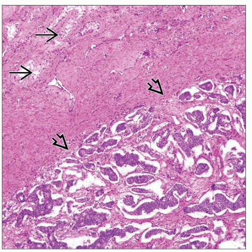 Testicular carcinoid tumor  shows a well-circumscribed nodule demarcated from the adjacent seminiferous tubules shows a well-circumscribed nodule demarcated from the adjacent seminiferous tubules  . The tumor has nested, trabecular, and insular growth patterns. . The tumor has nested, trabecular, and insular growth patterns. |
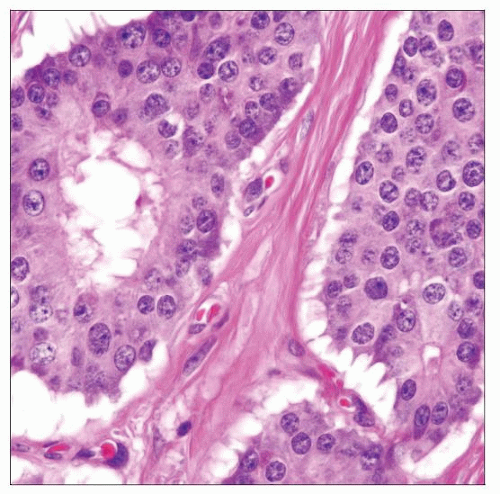 High-power view shows a carcinoid tumor with relatively uniform cells, round to ovoid nuclei, “salt and pepper” chromatin, and abundant eosinophilic and granular cytoplasm. |
TERMINOLOGY
Synonyms
Well-differentiated neuroendocrine carcinoma
Definitions
Well-differentiated tumor with neuroendocrine differentiation, which may occur as pure tumor, as component associated with teratoma or metastatic to testis
ETIOLOGY/PATHOGENESIS
Pathogenesis
Considered to be a monodermal form of teratoma
CLINICAL ISSUES
Epidemiology
Incidence
Extremely rare
Majority (70%) are pure carcinoid &/or associated with a teratoma (20%)
Rare cases of metastatic carcinoid (10%) from lung or gastrointestinal tract have been reported
Age
Range 10-83 years (average: 46 years); primary carcinoid: 44 years; metastasis: 61 years; carcinoid within teratoma: 38 years
In general, occurs in older age group than most other types of germ cell tumor
Presentation
Testicular enlargement ± pain
Equally distributed in left and right sides
May be associated with hydrocele (10%)
Carcinoid syndrome may occur (12%)
Laboratory Tests
5-hydroxyindoleacetic acid (5-HIAA) or metabolite of serotonin may be elevated
Stay updated, free articles. Join our Telegram channel

Full access? Get Clinical Tree


