Patient Story
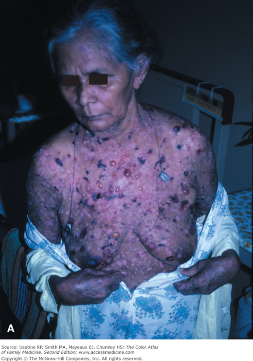
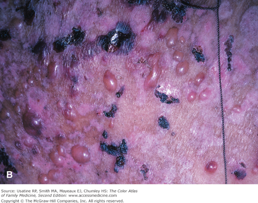
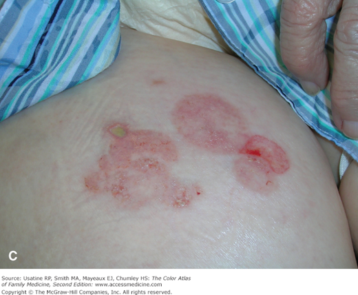
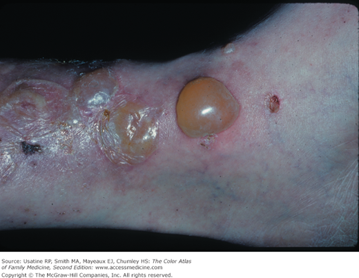
A native of Panama was seen for extensive bullous disease that is classic for bullous pemphigoid (BP) (Figure 184-1). The presence of numerous intact bullae would make pemphigus very unlikely. The patient was treated with oral prednisone and began to respond quickly. The patient eventually had a good outcome.
Introduction
BP is an autoimmune blistering disease of older adults that may cause significant morbidity and a poor quality of life. The term pemphigoid refers to its similarity to the blisters seen in pemphigus. However, BP is usually less severe than pemphigus vulgaris and is not considered a life-threatening condition.
Epidemiology
- BP is the most frequent autoimmune blistering disease of the skin (and mucosa).
- It typically affects persons older than 65 years of age but can occur at any age.
- There is no racial or gender predilection (a recent British population study, however, suggested an increased prevalence in women).1
- Its incidence may be on the rise.1
- Although it is not considered life threatening, it has been associated with an increased risk of mortality (hazard ratio [HR] 2.3, 95% confidence interval [CI] 2 to 2.7).1
Etiology and Pathophysiology
- BP is a chronic autoimmune disorder of the skin.
- Immunoglobulin (Ig) G autoantibodies against BP180 antigen of the basement membrane protein are considered pathognomonic and can be found in up to 65% of patients.2
- Anti-BP230 antibodies are present in virtually all patients but are not considered pathognomonic.3
- Binding of antibodies to the basement membrane activates the complement system, leading to chemotaxis of inflammatory cells (eosinophils and mast cells), which release proteases. The subsequent degradation of hemidesmosomal proteins leads to blister formation.
- There are several morphologically distinct clinical presentations:
- Generalized bullous form is the most common (Figure 184-1, 184-2, and 184-3). Tense bullae occur on both erythematous and normal-appearing skin surfaces. The bullae usually heal without scarring.
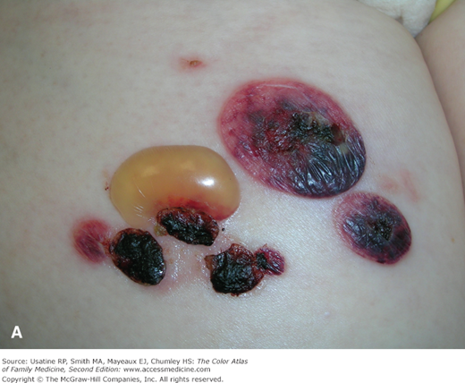
- Localized form of BP is less common and is limited to a small area of involvement (Figure 184-2).
- Vesicular (also known as “eczematous”) form is characterized by clusters of small tense blisters with an urticarial or erythematous base.
- Other forms are less common and include vegetative (intertriginous vegetating plaques), urticarial (without any bullae), nodular (resembling prurigo nodularis), acral (bullae on palms, soles, and face in children associated with vaccination), and generalized erythroderma (exfoliative lesions with or without vesicles/bullae).
- Generalized bullous form is the most common (Figure 184-1, 184-2, and 184-3). Tense bullae occur on both erythematous and normal-appearing skin surfaces. The bullae usually heal without scarring.
- Pemphigoid gestationis is a variant of BP that occurs during or after pregnancy. Lesions resolve after delivery, but may recur with subsequent pregnancies, or in the nonpregnant state (Chapter 76, Pemphigoid Gestationis).4
- Drug-induced BP has been reported with drugs containing sulfhydryl groups, including penicillamine, furosemide, captopril, and sulfasalazine.
Figure 184-2
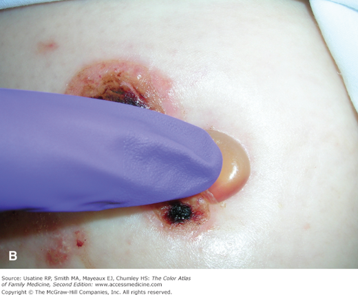
Localized bullous pemphigoid with large bulla on the thigh of this 91-year-old woman. Her biopsy for direct immunofluorescense demonstrated a linear band of igG at the dermal-epidermal junction. A. Bullae on the thigh. B. One week later there is a new bulla and the Asboe-Hansen sign is negative. C. One week later the bullae are healing as the bullous pemphigoid is being treated with clobetasol topically and the patient is taking doxycycline and niacin orally. (Courtesy of Richard P. Usatine, MD.)
Diagnosis
- Tense blisters that involve normal or inflamed skin or mucous membranes (Figures 184-1 and 184-2).
- Development of bullae is typically preceded by a prodromal phase characterized by intense pruritus with or without excoriations and eczematous (or urticarial) lesions. This phase can last for months, making early diagnosis difficult.5
- Nikolsky sign (wrinkling and sheet-like peeling of the skin when lateral pressure is applied to unblistered skin) is usually negative.6 Asboe-Hansen sign will be negative too. Bulla will not extend to surrounding skin when vertical pressure is applied (Figure 184-2B).
- Flexure surfaces of the arms and legs.
- Lower abdomen and groin.
- Mucous membranes are involved in 10% to 25% of cases.
Biopsy is required for establishing diagnosis and to differentiate BP from other conditions that can have a similar clinical presentation:
Stay updated, free articles. Join our Telegram channel

Full access? Get Clinical Tree


